VIENNA - CT has long been the go-to modality for the diagnosis of acute stroke, and its benefits are many. But MRI shows growing promise as an effective alternative, past ECR president Prof. Paul Parizel told attendees.
"Noncontrast CT and CTA are the cornerstones of stroke management, but technically, there's not much optimization that we can do with them," said Parizel, the David Hartley chair of radiology at the University of Western Australia in Perth, during a session on 1 March. "MRI has the potential to change management if it can be made more readily available."
Stroke imaging is optimized by effective protocols, and these are about "providing the diagnostic information or the endovascular treatment that matches clinical management," he noted, citing a 2001 paper published in the American Journal of Neuroradiology which outlined the "four Ps" of stroke assessment that remain pertinent today:
- Parenchyma: Rule out hemorrhage and assess early signs of acute stroke.
- Pipes (vessels): Assess extracranial and intracranial circulation for intravascular thrombus.
- Perfusion: Assess cerebral blood volume and flow.
- Penumbra: Assess tissue at risk of dying if ischemia continues.
So what's the best imaging tool for making these assessments? Parizel offered session attendees an overview, emphasizing that imaging must answer three questions for patients experiencing stroke:
- Is there an intracranial hemorrhage that contraindicates intravenous tissue plasminogen activator (tPA) treatment or endovascular therapy (EVT), or is there a large, well-established hypoattenuating infarct?
- Is there a proximal large vessel occlusion that can be treated with EVT?
- Is there a large core infarct seen at diffusion-weighted imaging, at CT angiography (CTA) of collateral vessels, on CTA source images, or at perfusion CT that is a relative contraindication to intravenous tPA or EVT?
Nonenhanced CT (NECT). NECT remains the first-line imaging modality for neurologic emergencies due to its speed, accuracy, and availability, Parizel said. It is used to exclude intracranial hemorrhage and identify large infarcts (i.e., those that put more than one-third of the brain at risk). Unfortunately, NECT has a "limited role in predicting and identifying early stage infarction, despite improvements in technique and analysis," he noted.
CT angiography (CTA). CTA is the best way to identify large vessel occlusion (LVO) and thus to expedite stroke treatment with endovascular therapy -- although Parizel did emphasize that starting treatment with intravenous tPA takes priority over advanced CT imaging in stroke patients.
Perfusion imaging. This technique can assess a patient's viable brain tissue and depict collateral circulation -- which is an important outcome predictor, he noted.
MRI. MRI shows promise beyond CT for identifying stroke, according to Parizel. (In fact, at the ECR meeting Japanese researchers reported that a 7-minute MRI protocol can now play a key role in the management of acute ischemic stroke patients.) Its advantages over CT include its lack of radiation, its sensitivity to acute ischemia, and its specificity in measuring the core volume of the infarction. MRI does have a few disadvantages, Parizel conceded, such as limited availability and potentially delayed workflow due to the need to screen patients for contraindications. But its benefits are compelling.
"Short protocol emergency brain MRI after negative noncontrast head CT is a cost-effective strategy in selected neurological patients with mild and unspecific symptoms, [and results] in lower costs and higher QALYs [quality-adjusted life years]," he said.
Even better, the development of portable MRI makes the modality available for point-of-care imaging -- although the image quality needs further development, according to Parizel.
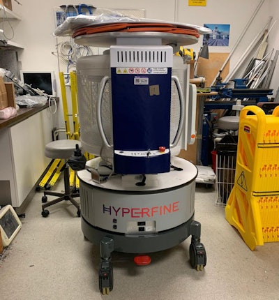
"At Royal Perth Hospital, we've acquired a mobile ultralow-field MRI scanner as part of a nation-wide research project sponsored by the National Imaging Facility (NIF), with a generous contribution from the hospital," he said.
 This ultralow field MRI scanner arrived recently at Royal Perth Hospital (RPH). This photo was taken in the store room before the machine went through the intake process of the hospital. RPH obtained the scanner as part of the PoCeMR (point-of-care MR) scheme funded by the National Imaging Facility in Australia. "The big question is whether the quality will be sufficient for clinical decision-making. Only the future will tell," commented Prof. Paul Parizel, who provided this photo.
"The purpose is to explore whether this technology can be used a mobile ultra-low-field MRI that can be used in point-of-care settings such as the intensive care unit, the emergency department, mobile stroke units, and resource-limited locations," he continued. "The image quality is not good -- field strength is 0.064 tesla -- but [still, portable MRI] could be part of the solution."
The bottom line for imaging and stroke management is providing timely information to clinician peers that integrates with stroke patient management, Parizel concluded.


