VIENNA - An automated software algorithm can be used to quantify emphysema on MDCT studies, offering radiologists a useful tool for diagnosis and follow-up of chronic obstructive pulmonary disease (COPD), according to research presented on Thursday at ECR 2017.
A team of researchers from Spanish image analysis software developer Quibim and La Fe Health Research Institute in Valencia, Spain, has developed an automated method that can segment and provide quantitative evaluation of emphysema by thirds of the lung, yielding better understanding of emphysema etiology.
"Automated lung emphysema quantification can help the diagnosis and follow-up of COPD," said Irene Mayorga-Ruiz of Quibim. She shared the research during a scientific session on Thursday morning.
Emphysema
Emphysema is a type of chronic obstructive pulmonary disease (COPD). Seeking to facilitate a recent paradigm change from qualitative to quantitative assessment of the disease, the Spanish group developed an automated algorithm that can segment, quantify, and characterize areas of emphysema, lung parenchyma, and blood vessels, Mayorga-Ruiz said.
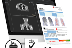
After the MDCT image is uploaded to the algorithm, the image data are converted to Hounsfield units and the image is then binarized. Next, the emphysema is segmented. In the study, the researchers evaluated two different segmentation methods: a fixed threshold of -950 HU and also a patented adaptive thresholding technique from Quibim. The group's method applies a specific threshold for segmenting emphysema to reduce bias caused by the patient's individual lung volume and the type of CT scanner used for image acquisition, she said.
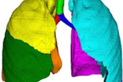
After extracting the lung and blood vessels, the software then classifies and labels the image with right and left lines as well as by different thirds of the lungs. Finally, the software quantifies the percentage and total volume of emphysema.
The researchers tested the software on MDCT scans from 39 patients: 22 men and 17 women. All images were acquired with a tube voltage of 160 kVp, tube current of 250 mA, slice thickness of 2 mm or less, and pixel size of 1 mm or less.
In testing, the emphysema quantification method takes 30 to 45 minutes on average, depending on the study size, she said. In addition, the software found that the right-lung volume was 8.7% higher for male patients and 9.5% greater for female patients, compared with left-lung volume.
Adaptive thresholding
Importantly, the algorithm's adaptive thresholding quantification of emphysema was 50% less, on average, than the fixed thresholding technique, indicating a superior performance in characterizing emphysema, according to the group.
"The adaptive thresholding, due to the [image-specific air threshold], is able to perform a better characterization of emphysema," she said.
The software's ability to segment and quantify emphysema by each third of the lung enables clinicians to infer the etiology of the emphysema, Mayorga-Ruiz said.
Of the 39 patients, 29 were diagnosed with emphysema caused by external agents, as the emphysema was mainly detected in the apex of the lung on the software, according to Mayorga-Ruiz. An additional two patients presented as having the genetic disorder alpha-1-antitripsine deficiency; the emphysema was detected mainly in the base of the lung in these patients. Eight patients had nonspecific findings due to the percentage of emphysema being below the normal range of 5%, she said.
The team is now evaluating the emphysema segmentation algorithm with low-dose CT, Mayorga-Ruiz said.


