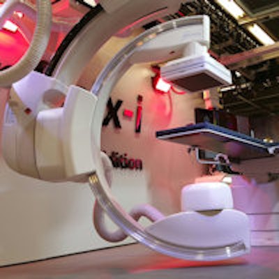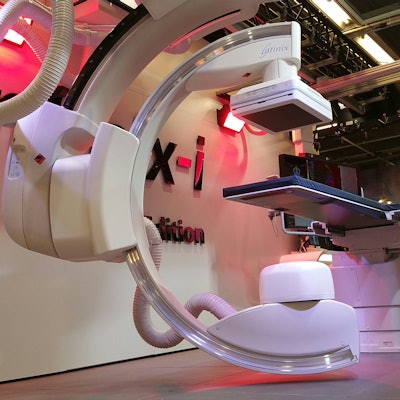
VIENNA - Toshiba Medical Systems is taking advantage of this week's ECR 2016 to launch Infinix-i Rite Edition, a new interventional x-ray system that features a dual C-arm design for faster and more efficient operation and high-speed 3D studies.
Toshiba designed Infinix-i Rite Edition as a ceiling-mounted flat-panel digital system with one C-arm positioned inside the other. Both C-arms can move independently, enabling the system to perform a 210° rotation very quickly, at a rate of 80° per second, according to the company. This improves image resolution and can reduce the need for contrast material.
In addition, the C-arm can flip and rotate in any direction, and the flat-panel detector can be placed underneath the patient; thanks to the ability to move the C-arm laterally, the entire system can be moved away from the patient during procedures.
 Infinix-i Rite Edition features a novel dual C-arm design.
The design of Infinix-i Rite Edition gives the system flexibility in performing advanced 3D studies, such as a complete head-to-toe interventional procedure. In addition, the unit's 30 x 40-cm digital detector is automatically synchronized with the beam collimator.
Infinix-i Rite Edition is shipping now in Europe, and a couple of systems are in operation in Japan. The system is not yet available in the U.S.
In other ECR 2016 news, Toshiba is giving congress delegates a look at OrthoMod 3D, a new spinal imaging technology designed for use with x-ray. OrthoMod 3D consists of an optical imaging system mounted on a small cart that can be positioned next to a bucky-based x-ray system, such as Toshiba's Ultimax-i unit.
The unit uses a high-resolution optical camera that captures 3D images of back morphology that can be matched to x-ray images -- previously, patients had to be referred to other modalities such as MRI for 3D information. The technique can give physicians a better look at whether patients have spinal conditions that involve rotations and twists in the spine, such as scoliosis.
OrthoMod 3D was developed by AXS Medical, a division of French medical technology developer DMS-Apelem, and Toshiba is the official distributor of the system. The unit is currently shipping in Europe; it is not yet available in the U.S.
In CT, Toshiba is demonstrating a reconstruction computer dedicated to supporting the use of model-based iterative reconstruction (MBIR) for radiation dose reduction on its Aquilion One CT scanner. The computer enables MBIR studies to be created in as few as three minutes, reducing radiation dose by up to 80% and improving image quality. Available as an option for Aquilion One, the computer is now shipping after first being launched at RSNA 2015.
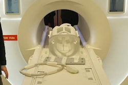
In MRI, Toshiba is reprising the novel heads-up display system for patients that it demonstrated last year in Chicago. The system uses a semitransparent dome screen to project soothing nature scenes inside the bore of the MRI scanner to counter patient claustrophobia, and it could potentially also be used for functional MRI research.
Toshiba sees the system as a visual counterpart to its Pianissimo technology, which it introduced years ago to reduce noise inside the MRI scanner. Using both systems together could counteract the feelings of claustrophobia and discomfort that some patients experience, which can lead to canceled exams. The display is being shown as a work-in-progress and is not currently shipping.
Toshiba is also discussing its effort to reduce MRI total cost of ownership, such as with its Vantage Elan 1.5-tesla scanner. The scanner features zero cryogen boil-off, and Toshiba believes that users can save 250,000 euros to 500,000 euros in running costs over the seven- to eight-year life of a scanner.
In the ultrasound section of its booth, Toshiba is discussing the Innovation 2016 upgrade for its Aplio Platinum line of scanners, with a special focus on its Superb Micro-Vascular Imaging (SMI) protocol. The SMI protocol allows clinicians to visualize smaller vessels in and around structures like tumors, inflamed tissue, and lymph nodes, thanks to its capacity for visualizing low-velocity flow at a level far beyond the capability of conventional Doppler techniques.


