As the dust begins to settle on ECR 2015, now is a great time to reflect on the meeting and identify the major trends affecting medical imaging. Central themes circulating the halls of the Austria Center Vienna included the unification of European radiology, as well as teleradiology and outsourcing, radiation dose monitoring, digital breast tomosynthesis, and hybrid imaging.
No. 1: European radiology unites at ECR
Europe came together in near-perfect harmony at ECR 2015. People of different nationalities mixed, interracted, and shared experiences in a natural, positive, and spontaneous way. The overwhelming mood was, "What I can I give to the congress?" not "What can I take from it?"
 Dr. Bernd Hamm has seen both Berlin and the Charité transformed over the past 20 years.
It's easy to forget that European radiology has not always been one big, happy family. ECR got off to a decidedly dodgy start when it first moved to a new permenant home in Vienna in 1991. The French and the Brits stayed away for years, preferring to attend their national meetings and the RSNA show. The continent's leading research centers continued to target Chicago, sending their big hitters and A-listers across the Atlantic every November and dispatching their juniors and also-rans to the Austrian capital.
How things have changed for the better, and Congress President Dr. Bernd Hamm personifies this evolution. Just as his home city of Berlin has emerged united, resurgent, and purposeful, so has his institution, the Charité, as well as the ECR. The dark days of the Cold War are long forgotten.
ECR 2015 was not utopia, of course. The plastic yellow security wristbands were unpopular, and too many sessions on related topics were held concurrently and in the wrong-sized rooms. But overall, the congress is alive and well and continues to thrive, and it improves each year. It has become a fine showcase for European radiology and something to be proud of. Can we really expect any more than that?
No. 2: Teleradiology, radiation dose monitoring, decision support, & mobile devices
A wide range of imaging informatics topics took center stage at ECR 2015, as teleradiology, radiation dose monitoring, clinical decision support, and mobile devices drew considerable interest among attendees.
Debate is intensifying over how best to balance the acknowledged benefits of teleradiology with the technology's danger for facilitating the commoditization of radiology. Despite having concerns over its potential risks, most Italian radiologists and French radiology residents were still inclined to hold a positive view of teleradiology, according to the results of surveys presented at the meeting.
Presentations on radiation dose monitoring featured heavily in the program, as attendees sought to learn from the experience of trailblazing institutions in monitoring and analyzing patient radiation dose metrics. Also, clinical decision support entered a new era in Europe with the official launch at ECR 2015 of ESR iGuide, a collaboration between the ESR and U.S. software developer National Decision Support Company (NDSC). ESR and NDSC plan to roll out iGuide in phases; pilot sites in all the major European sites are slated to come online in the third quarter.
Issues surrounding the utilization of mobile devices in radiology continue to generate great interest. Among the key topics included the importance of protecting patient privacy when using these devices for remote diagnosis, as well as the need for guidelines to direct their appropriate utilization.
 Figure 1's automatic face-blocking feature detects faces and blocks them, and the manual block feature allows a user to quickly and easily block anything else that might identify a patient.
Speaking of patient privacy, social media technology -- not usually thought of as a secure way to share patient information -- is being developed to enable radiologists and other physicians to securely and privately share information such as medical images without identifying patients. One such tool is Figure 1, which can be downloaded for free from both the iPhone App Store and Google Play.
No. 3: CT expands role as prognostic tool
Long the workhorse modality in medical imaging, CT shows no sign of slowing down. At ECR 2015, numerous scientific sessions demonstrated CT's growing role as a prognostic tool for diseases such as stroke and heart attacks.
For example, Dutch researchers demonstrated that almost 20% of middle-aged athletes are at risk of coronary events, even if they have low Framingham risk scores and little evidence of ischemia. The group performed coronary calcium scoring on 314 athletes older than 45, finding that 19% had coronary artery disease, and 5% had coronary stenoses greater than 50%. The issue is serious enough that the researchers believe CT screening exams could be warranted for middle-aged athletes. The number needed to screen to prevent one cardiovascular event compared favorably with other screening tests such as mammography, they added.
 Images are of a study participant with no coronary artery disease (right) and a second participant, his brother (left), who has obstructive coronary disease. Both are cyclists. Images courtesy of Dr. Thijs Braber.
A further study pointed to CT's ability to tell when patients who are suspected of pulmonary embolism actually have pulmonary hypertension instead. Israeli researchers used CT angiography (CTA) and an investigational software application to automatically measure cardiac chambers. They developed a set of parameters that enabled them to differentiate the two conditions with 90% accuracy compared with echocardiography.
Another automated software-based tool fell short, however, compared with manual techniques in determining the extent of stroke on perfusion CT scans. Italian researchers found that maps of perfusion abnormalities generated via a software tool had lower sensitivity than perfusion maps drawn manually.
As has been the case in recent years, concerns about CT radiation dose were an urgent priority. Fortunately, academic researchers and vendors demonstrated progress in addressing this issue, with a number of courses on managing and tracking radiation dose, as well as updates on the EuroSafe radiation safety campaign, first launched at ECR 2014.
No. 4: Digital breast tomosynthesis attracts growing attention
It was definitely a case of tomo, tomo, everywhere. Digital breast tomosynthesis (DBT) proved to be incredibly popular at ECR 2015. Delegates packed each session that discussed DBT to learn what's new with the modality.
Mainly, the trend seems that DBT is edging its way toward becoming a primary screening modality instead of an adjunct to mammography, a day DBT advocates long for.
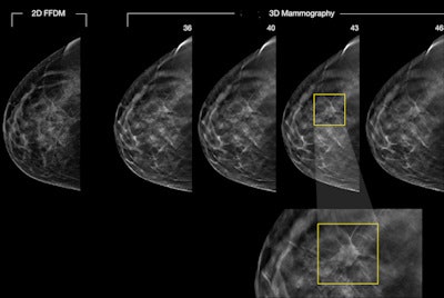 A 46-year-old woman's routine screening mammogram: 2D mammogram is essentially negative. Digital breast tomosynthesis (3D mammography) images reveal a 15-mm spiculated mass between slices 38-48, diagnosed as invasive ductal carcinoma, grade 2. Figure courtesy of Dr. Per Skaane, PhD.
This was especially evident at an industry symposium. Dr. Sarah Friedewald from the Lynn Sage Comprehensive Breast Center in Chicago discussed how 90% of her facility's screening patients are imaged with tomosynthesis. Dr. Marina Alvarez Benito from the Reina Sofia Hospital in Cordoba, Spain, emphasized that while DBT adoption initially faced some obstacles, now it's gaining traction -- in the last year, more than 50 tomosynthesis systems have been installed in her country. She and her colleagues are conducting a prospective study to evaluate the validity of tomosynthesis plus synthesized mammography exams compared with digital mammography as a gold standard in a screening program.
Many studies at ECR 2015 focused on synthesized mammography, or constructing 2D mammograms from DBT datasets using image-processing software. Several researchers have begun advocating the use of synthesized 2D images plus DBT, stating that they work as well as conventional 2D mammography plus DBT. Researchers have also continued to compare DBT with mammography in detecting calcifications and study the technology's impact on recall rates, masses, and more.
One thing's clear: Evidence continues to mount that DBT trumps mammography in many regards.
No. 5: Radiology realizes the future is about hybrid scanners
Hybrid imaging was a central theme at ECR 2015. In addition to some excellent scientific sessions and the honorary lecture by Dr. Gerald Antoch from Düsseldorf, Germany, there were basic and advanced training courses -- something for everyone.
Uncertainty still surrounds the precise clinical applications of PET/CT, and the jury is still out on whether PET/MRI can ever be cost-effective, but it's a positive development that radiology is at least starting to address the key questions in hybrid imaging.
 The Swedish president of ECR 2016, Dr. Katrine Åhlström Riklund, PhD, is a radiologist who is also licensed in nuclear medicine.
Antoch is convinced that the hybrid imager must be a specialist, both in radiology and nuclear medicine, and he thinks doctors need to be trained in both disciplines to read hybrid datasets autonomously. But is such an approach practical and workable? Which type of training system can work best across Europe? Can radiology and nuclear medicine work together more closely in this area? And how can the turf battles inherent in hybrid imaging be avoided?
Ironically, the European Society of Radiology (ESR) and the European Association of Nuclear Medicine (EANM) are both based in Vienna, and some former ESR staff members have moved across to the EANM, but the two organizations rarely seem to communicate with each other. If radiology and nuclear medicine are going to collborate in the future in a meaningful way, then this situation must change.
Hybrid imaging looks set to move up the agenda even more at ECR 2016, not least because the Swedish Congress president, Dr. Katrine Åhlström Riklund, PhD, is a radiologist who is also licensed in nuclear medicine. Watch this space ...


