The landscape of molecular imaging has changed over the past two years, with advances having been made in image-guided drug delivery, gene-based therapy, and cellular therapy. For the latter, cell-labeling methods and image-based magnetic control of cells now enable the monitoring and guidance of therapy. Potential future applications, such as intracellular heating, could place imaging at the very center of therapy delivery.
A panel of experts will show, in a dedicated minicourse today at the European Congress of Radiology (ECR), that molecular imaging can -- and soon could -- offer improvements in the efficiency of treatment.
The recent development of cell-labeling techniques using superparamagnetic nanoparticles has confirmed the role of imaging in monitoring cellular therapies. In particular, researchers have been able to develop high-resolution cellular imaging, using cryoprobes in MR applications. This combination enables high-resolution imaging with a sharp definition of cells in a given organ.
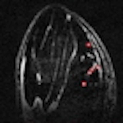
 High-resolution 3D MR image of (A) axial, (B) sagittal, and (C) coronal slices of the calf muscle of a mouse treated with M0 macrophages subpopulations at 24 hours postinjection, showing signal voids that are punctual (white arrows) and do not correspond to vessels. (D) Example of the processed image, which allows the quantification of hypointense punctual regions. (Figure provided by Prof. Olivier Clément)
"It's probably the main discovery we have made over the past two years. By labeling macrophage cells in an ischemic pad of a rat's foot, we could clearly see the colonized cells and the ischemic territory," said Professor Olivier Clément, a radiologist working at European Hospital Georges Pompidou in Paris, France.
Projects such as the European Network for Cell Imaging and Tracking (ENCITE), coordinated by the European Institute for Biomedical Imaging and Research and funded by the EU (encite.org), have played a major role in this progress, the researcher underlined.
These techniques have shown no adverse effects on cell proliferation and functionalities while conferring magnetic properties on various cell types. The magnetic labeling of living cells creates opportunities for numerous biomedical applications, such as individual cell manipulation and magnetic control of cell migration.
Clément and his team are currently working on facilitating cell-homing in myocardial infarct cellular therapy, by placing an external magnet over the heart to create a local field gradient which induces magnetic targeting.
"We have been working on a study to improve cell-homing in the heart and succeeded in having more cells when using a magnet. We still haven't been able to show a therapeutic effect with this technique, but other studies have, and they could show a long-term effect, including scar reduction," he said.
Intracellular heating will, probably soon, become a field of investigation for many researchers. With this method, one will be able to kill cancerous cells and tumors or liberate drugs contained in a liposome by redirecting the heat, created by the use of magnetic fields, to superparamagnetic nanoparticles.
"Let's imagine that we have a liposome containing drugs and superparamagnetic nanoparticles. With a magnet, you can drag the liposome exactly where you want it; to a tumor, for instance. Then, you just have to heat your liposome, using external microwaves, after which it will break and allow for chemotherapy. This is still a concept, but it could become a third application in the short term," Clément said.
The transition of these techniques into clinical practice remains a long-term prospect. Many things are still to be done, including toxicity tests. On the bright side, cellular therapies such as blood transfusion and bone marrow transplant already work without image-based monitoring.
Molecular imaging should, however, prove very useful for tissue transplants and regeneration by showing exactly what happens.
"It is not clear at all whether the injected cells will replace organ function, for instance in myocardial failure. A cell can also trigger signals that help the heart to function better. In this case, imaging helps us understand exactly what happens," said Clément, who foresees its upcoming clinical application in the heart.
"Imaging can significantly improve the accuracy of treatment," said Dr. Michal Neeman, a researcher working on the development of reporter genes for MRI at Weizmann Institute in Rehovot, Israel.
Reporter genes encode proteins that can be detected by imaging and serve as a surrogate for the activity of a particular promoter area. For example, reporter genes are used to follow the activation of tumor-associated fibroblasts as they penetrate tumors, and follow the expression of angiogenic and lymphangiogenic growth factors by tumors.
Different reporter genes have been developed to allow detection by different imaging modalities. The most common reporter genes are the fluorescent and bioluminescent proteins that emit light and can be detected using sensitive cameras. Reporter genes are now also available for nuclear imaging and MRI.
"I believe the next steps will be to prove efficacy and safety in specific diseases," Neeman concluded.
The first success story is the treatment of hemophilia B, as shown by a study recently published in the New England Journal of Medicine by researchers at University College London and St. Jude Children's Research Hospital in the U.S.
Originally published in ECR Today March 5, 2012.
Copyright © 2012 European Society of Radiology


