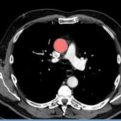
Medical imaging investigations, which consist almost entirely of 2D scans, are shortchanging radiology students in a 3D world where most real-life clinical imaging is displayed in volumetric images, according to researchers from the Netherlands.
A group from the University Medical Center Utrecht wanted to know how well 246 medical students could use a set of 2D images they were shown to answer related 3D volumetry questions. A subset of them also took a human cadaver anatomy test. The results showed that human cadaver test scores weren't correlated with 2D image scores, but volumetric image (VI) scores were (r = 0.44, p < 0.05).
"We found that volumetric image test scores correlated significantly with the external measure, while 2D image test scores did not," wrote lead author Dr. Cécile Ravesloot in an email. "Reliability estimates for volumetric CT image scores were higher than for 2D CT image scores, and students considered volumetric image questions to be more representative of the educational program and clinical practice."
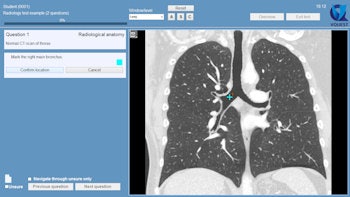
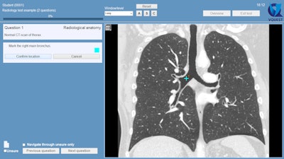
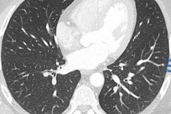
Example of an "indicate question" in VQuest. The student is asked to mark an anatomical structure in the volumetric image. Students can scroll through the image, change contrast settings manually or with the preset window/level menu, or change viewing direction by clicking the buttons (A, S, and C). The teacher decides which manipulation tools the students are allowed to use during the test or for a particular question. All images courtesy of Dr. Cécile Ravesloot.
Innovation now the norm
Acquiring basic radiological knowledge and image interpretation skills for medical students is an increasingly important mission as diagnostic imaging has become a prominent tool in daily clinical practice, noted Ravesloot, a radiology resident and PhD student in radiology education, and her Utrecht colleagues Dr. Anouk van der Gijp, Marieke van der Schaaf, PhD, et al (Academic Radiology, 12 February 2015).
Rather than looking at tiled image slices as in the past, today the use of innovative image displays is now the norm, the study team wrote.
"This allows the radiologist to scroll through 3D datasets (stack viewing), adjust window level, and use advanced image reconstruction tools, such as on-the-fly multiplanar reformatting," they wrote.
The data for a single cross-sectional exam can involve hundreds of slices, which observers can scroll through in various planes and contrast settings. A vast amount of visual information must be processed and interpreted by the observer, the researchers explained, and because image interpretation has changed so radically, radiology education should change as well.
To that end, the study team evaluated a new method for testing medical students' radiological anatomical image interpretation skills using stacks of CT slices (volumetric images), and compared it with the traditional single slices or 2D image testing.
Testing the difference
The test was made up of 75 questions, including 40 CT anatomy questions consisting of 20 questions based on 2D images and 20 based on volumetric images. As there is no gold standard for anatomy interpretation skills, the researchers decided to use a human cadaver anatomy test, which was taken by 33 participants; the researchers correlated scores on the cadaver test with those from the 2D and volumetric subtests to determine accuracy. Finally, the participants received a questionnaire about perceived representativeness and difficulty of the radiology test.
In all, 246 medical students completed tests; all of the test takers had completed a two-year radiology education program including basic radiological skills on common diseases as well as radiological anatomy as part of their preclinical training. Tests were administered using VQuest, a locally developed digital testing program that allows for stack viewing and authentic image manipulation.
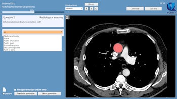
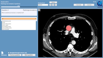
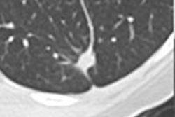
Example of a question in which students are asked to name the marked structure. In this case, they need to choose from a long list of options, which they can only open by typing at least two letters of their answer. All alternatives including these two letters are then shown, and the student can click on the preferred answer. This allows for automatic answer checking.
In the volumetric test questions, for example, the participants could change viewing directions by pushing buttons, for instance to view sagittal or coronal views, and after contrast administration they could alter the contrast settings (abdomen, bones, lungs) by choosing preset window levels.
3D imaging makes for easier learning
The results showed that mean 2D image scores were significantly higher than volumetric scores for test B participants (t [124] = 8.7; p < 0.001). Mean radiology test scores of human cadaver test participants (83.2%, standard deviation [SD] 8.0) were higher than for radiology test participants who did not volunteer in the human cadaver test (78.2%, SD 11.5). This difference in mean radiology test scores was significant for test A participants (t (119) = ≤ 2.4; p = .02).
Volumetric image test scores correlated significantly with the external measure (r = 0.44, p < 0.05), while 2D image test scores did not, Ravesloot wrote. In addition, reliability estimates for volumetric CT image scores were higher than for 2D CT image scores, and students considered volumetric image questions to be more representative of the educational program and clinical practice.
"Remarkably, students perceived the volumetric image as less difficult, but did not receive higher scores than for 2D image questions; indeed we found a significantly lower mean score in one group," Ravesloot noted. "Our results indicate that volumetric CT images in tests probably better reflect relevant skills and seem to yield higher test quality in different respects."
The results show that volumetric imaging contributes to the external validity and reliability of radiological anatomy testing, the authors wrote. Unlike in the 2D questions, volumetric questions correlated to an external measure of anatomy knowledge; i.e., the cadavers. The use of cadavers is certainly not the perfect 3D external measure, the authors noted, but because there is no gold standard, it is probably the best alternative available.
"[D]igital radiological VI interpretation in current clinical practice requires a more holistic 3D understanding of anatomy, which is better reflected by VI than 2D image radiology test scores," they wrote. Another study limitation is that only a small group of readers took the cadaver anatomy test.
Previous studies have demonstrated that teaching anatomic knowledge is important for radiology image interpretation. The results of the present study suggest that 3D "is particularly important for radiological image interpretation," the authors wrote.
"The results indicate that VIs improve radiological anatomy test quality on all studied quality aspects," Ravesloot et al wrote. Due to the rapid growth of imaging and the need for high-quality of volumetric image interpretation, volumetric images with stack viewing and multiplanar reformatting tools should be included in radiology tests, especially in radiological anatomy testing.
More research will improve volumetric image testing further, and the group is planning to conduct it, she added.
"We ... plan to do more studies on the quality of volumetric image testing on different expertise levels, e.g., radiology clerks and residents, and how the quality might be further improved," Ravesloot said. "We are currently investigating methods and question types that will provide teachers insight into gaps in the image interpretation process of learners. Ultimately, we strive to use these methods to develop feedback tools to stimulate image interpretation skill development."


