Using SPECT, researchers in France have detected functional abnormalities in certain regions of the brain in fibromyalgia patients. The findings support the idea that fibromyalgia symptoms are related to a dysfunction in the parts of the brain where pain is processed.
The purpose of the study, based at the Service Central de Biophysique et de Médecine Nucléaire in Marseille, was to investigate the specific clinical correlation of brain SPECT perfusion abnormalities reported in fibromyalgia, one of the most common causes of musculoskeletal pain and disability.
The lead author of the research, which is published in the November issue of the Journal of Nuclear Medicine (November 2008, Vol. 49:11, pp. 1798-1803), was Dr. Eric Guedj.
According to the National Institute of Arthritis and Musculoskeletal and Skin Diseases (NIAMS), fibromyalgia syndrome is a common and chronic disorder characterized by widespread muscle pain and fatigue. People afflicted with fibromyalgia also experience multiple tender points, such as on the neck, shoulders, back, hips, and upper and lower extremities, where they feel pain in response to slight pressure.
As many as one in 50 Americans have the syndrome, with women accounting for 80% to 90% of that population.
Patient enrollment
Researchers consecutively enrolled 20 women with fibromyalgia with a mean age of 48 years, ranging from 25 to 63 years, in the study. All patients underwent a general medical assessment by one investigator to confirm the diagnosis. The study excluded women who were pregnant and had a concomitant major medical or psychiatric disease.
The study also enrolled 10 healthy women in a control group to determine areas of significant hypo- and hyperperfusions in patients.
Patients received an injection of 740 MBq of 99mTc-ethylcysteinate dimer and were kept at rest for an hour in quiet surroundings with their eyes closed. SPECT was performed using a double-head rotating gamma camera (e.cam, Siemens Healthcare, Erlangen, Germany) equipped with a fanbeam collimator.
Researchers measured pain intensity with a visual analog scale, the Questionnaire Douleur de Saint-Antoine Scale (QDSA), and the Tübingen Pain Behavior Scale (TBS). QDSA measures different components of subjective pain, which describes the sensory and the affective dimensions. The higher the QDSA score, the worse the condition. TBS rates the presence of 11 pain behaviors, such as groaning, slow movements, and pain-based refusal to perform activities. It is based on a scale of 0 to 2, with 0 as none and 2 as always.
FIQ score
Disability was evaluated using the Fibromyalgia Impact Questionnaire (FIQ), which is designed to measure the severity of fibromyalgia disease symptoms. FIQ consists of visual analog scales and questions regarding limitations on daily living activities, and it evaluates the components of health status that are believed to be most affected by fibromyalgia.
FIQ scores range from 0 to 100, with 100 as the worst condition or most serious form of the disease. In the study's final analysis, patients with fibromyalgia had an average FIQ score of 66.6 (±12.9).
Researchers also noted that the FIQ total score was "positively correlated with bilateral parietal perfusion, including postcentral cortex. These clusters of correlation were included in the areas of significant hyperperfusion. FIQ total score was also negatively correlated with perfusion of a left anterior temporal cluster, included in the areas of significant hypoperfusions. No other clinical correlation was observed with regional cerebral blood flow."
 |
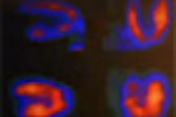
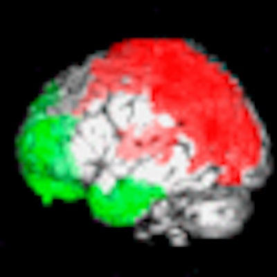
| The images show the anatomical localization of peak significant differences between brain SPECT of patients with fibromyalgia and healthy subjects. Patients with fibromyalgia exhibited posterior hyperperfusion (red), including of the somatosensory cortex, and hypoperfusion (green) of frontal, cingulate, temporal, and cerebellar cortices. Images courtesy of the Journal of Nuclear Medicine. |
Compared with healthy controls, patients with fibromyalgia exhibited posterior hyperperfusion, including of the somatosensory cortex, and hypoperfusion of the frontal, cingulate, temporal, and cerebellar cortices in particular, the temporal hypoperfusion including the polar and mediobasal cortices.
The study concluded that brain perfusion abnormalities in patients with fibromyalgia "are independent of the patient's anxiety and depression status and correlate with the clinical severity of the disease, expressed by the disability and evaluated by the FIQ total score."
Because FIQ has been suggested as "the most logical measure to evaluate functional status in clinical trials in fibromyalgia syndrome," the authors continued, "these findings reinforce the concept of using brain perfusion SPECT to objectively evaluate therapeutic results in controlled studies."
Guedj and colleagues also noted in the study that disability is frequently used in controlled clinical trials to evaluate treatment response. Because molecular imaging techniques such as SPECT can help predict a patient's response to a specific treatment and evaluate brain-processing recovery during follow-up, SPECT could prove useful when integrated into future pharmacological controlled trials.
By Wayne Forrest
AuntMinnie.com staff writer
November 7, 2008
Related Reading
Receptor availability may explain poor opioid response in fibromyalgia, October 29, 2007
Brain scans document fibromyalgia pain, June 18, 2002
Copyright © 2008 AuntMinnie.com