The promise of a diagnostic advantage in mammography using powerful 7-tesla MRI was underlined by preliminary results presented at the 2009 European Congress of Radiology (ECR). Although image quality was improved compared to 1.5-tesla MRI, using such a powerful system in a clinical environment might be problematic due to current patient safety rules.
First results from an early-stage, 17-patient German study, led by Dr. Lale Umutlu of Essen University Hospital, revealed a distinct diagnostic advantage to using 7-tesla versus 1.5-tesla MRI in the delineation of detailed anatomical and pathological breast tissue.
The objective was to generate a specific MR mammography protocol for a 7-tesla whole-body scanner and compare the results with gold standard 1.5-tesla MRI.
Using a single-loop transmit/receive coil (Rapid Biomedical, Würzburg, Germany) with a 7-tesla whole-body MRI system (Magnetom 7T, Siemens Healthcare, Erlangen, Germany), the researchers scanned 10 healthy volunteers and seven patients with known breast cancer.
Images were generated using two sequences on the Magnetom 7T system: T2-weighted turbo spin-echo (TSE) and dynamic contrast-enhanced T1-weighted spoiled gradient-echo (3D-FLASH).
All patients were rescanned on a 1.5-tesla whole-body MRI system (Magnetom Espree, Siemens Healthcare) within 48 hours, using a standardized clinical protocol. Subtraction images were acquired at both 1.5-tesla and 7-tesla field strengths (shown below).
Results indicated that at equivalent acquisition times, the 7-tesla scans presented more detailed anatomy and depicted 100% of known lesions in the dynamic T1-weighted images.
"We were able to transform the higher signal-to-noise ratio generated by the 7-tesla magnet into higher spatiotemporal resolution. This provides a better delineation of small anatomical structures of the fibrotic tissue as well as the lesions," Umutlu said. "Conclusively, the radiologist can depict and evaluate the size, margins, and morphology of suspicious breast lesions with greater precision."
 |
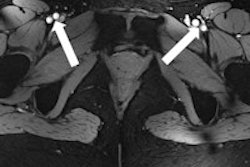
| T1-weighted postcontrast imaging of a 49-year-old woman; suspicious lesion in the lateral side of the breast at 1.5 tesla (A) and more clearly at 7 tesla (B). All images courtesy of Dr. Lale Umutlu. |
When asked about the potential to improve diagnosis in the clinic or even change care for breast cancer patients, Umutlu suggested that "if possible satellite lesions or a filtration of the pectoral muscle were to be depicted due to the higher spatial resolution at 7 tesla, that might induce a change in treatment plans."
 |
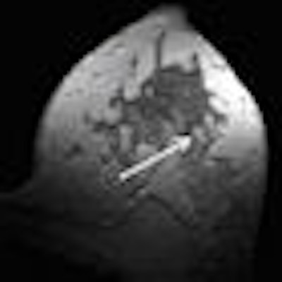
| Subtraction imaging of the same patient. In comparison to 1.5 tesla (A), the suspicious lesion on the lateral side of the left breast (broad arrows) shows brighter contrast enhancement at 7 tesla (B); evaluation of lesion size, margins, and morphology are improved at 7-tesla imaging, whose more explicit contrast enhancement allows the depiction of further satellite lesions (dashed arrow). |
No differences were apparent between the T2-weighted results generated at 1.5-tesla versus 7-tesla, with respect to their clinical value.
Limitations and potential
The results suggest a possible future role for 7-tesla MRI in breast cancer detection and fine detail characterization. But such ultrahigh-field imaging in the clinic is somewhat restricted at present by safety regulations governing specific absorption rate (SAR), the rate at which radiofrequency (RF) energy is absorbed by the body.
SAR ratings reflect the potential for heating biological tissue as the pulsed RF energy required to elicit MR signals is absorbed. While a fundamental element in the creation of every MR image, it is notable that doubling the magnet field strength, even from 1.5-tesla to 3-tesla, quadruples the SAR.
Concerned by the risk of generating hotspots during MR imaging using such powerful magnets, SAR limits are enforced by the U.S. Food and Drug Administration and other healthcare safety groups. Umutlu hopes the development of more advanced coil concepts will in time help to address the issue.
"We believe that 7-tesla breast MRI has high diagnostic potential, but it now needs to be evaluated in intraindividual comparison studies after the implementation of dedicated breast coils," Umutlu said.
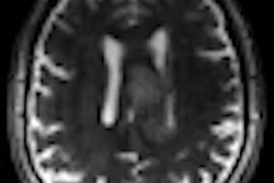
In related work, a separate study on brain imaging published by researchers at Essen University Hospital suggested that the higher diagnostic sensitivity of 7-tesla MRI was successful in detecting multiple sclerosis lesions.
By Rob Skelding
AuntMinnie.com contributing writer
March 13, 2009
Related Reading
MR features help radiologists identify triple-negative breast cancer, March 10, 2009
Joint Commission may survey for MRI safety, March 5, 2009
7-tesla MRI pinpoints ankle damage in marathon runners, March 8, 2008
MRI highly sensitive in breast cancer detection, January 9, 2008
Copyright © 2009 AuntMinnie.com