Coronary CT angiography (CTA) and MR myocardial perfusion imaging (MPI) work best as a team for evaluating chest pain patients with suspected coronary artery disease, according to a new study published online in Radiology.
The 145-patient comparison confirmed that while coronary CTA is excellent for ruling out disease, MR retains an edge in assessing the hemodynamic significance of lesions.
While coronary CTA has emerged as a valuable way to exclude stenosis, few studies to date have included many patients at low-to-intermediate risk of coronary artery disease (CAD), wrote Dr. Jan G. J. Groothuis and colleagues from VU University Medical Center in Amsterdam, Netherlands.
MR MPI, for its part, "has high spatial resolution and is thus able to demonstrate even small, subendocardial perfusion defects," and is "at least as accurate" as SPECT in demonstrating CAD. And as MR delivers no radiation, it "may serve as the ideal additional functional imaging technique" for these patients in tandem with coronary CTA, the researchers wrote (Radiology, January 20, 2010).
The study compared 64-detector-row CT (Sensation 64, Siemens Healthcare, Erlangen, Germany) with 1.5-tesla MRI (Sonata/Avento, Siemens Healthcare) in patients with chest pain and a low-to-intermediate probability of heart disease. There was a mean 4.6 days (± 3.0) between the exams.
In all, 148 consecutive chest pain patients (45% men, mean age 57 years ± 10 years) were analyzed for the study, after exclusion of candidates with a history of coronary artery disease, arrhythmias, renal insufficiency, allergies to contrast, etc.
Beta-blockers and nitroglycerine were administered before imaging; thereafter, the maximum heart rate required for inclusion was 65 bpm or fewer. The mean pretest probability of disease was 36% ± 20%, median 27%.
Coronary CTA images were acquired at 64 x 0.6 collimation, 370-msec rotation, 900 mAs, 120 kV, and flying z-focus after contrast administration (Ultravist 300, Bayer Schering Pharma, Berlin), injected at 5 mL/sec, followed by a saline flush. Data were analyzed by consensus of a radiologist and a cardiologist.
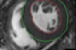
MR MPI was performed using an eight-element phased-array cardiac receiver coil. All images were acquired with ECG triggering and breath-holding. First-pass myocardial perfusion was assessed using a dynamic single-shot saturation recovery gradient-echo planar pulse sequence (repetition time echo-planar imaging factor of four, 160 x 144 matrix, 2.5 x 2.5 x 10-mm voxel size) and accelerated by parallel imaging using time-adaptive sensitivity coding, the authors reported. Gadolinium-based contrast (Magnevist, Bayer Schering Pharma, 0.1 mmol/kg body weight) was followed by a saline flush.
The epicardial fat signal was suppressed; three left-ventricle short-axis sections were acquired for every heartbeat. Stress imaging was begun three minutes after adenosine (140 μg/kg per minute). Data were analyzed, image quality graded, and diagnosis was made by consensus of two experienced cardiologists blinded to the coronary CTA data.
Two modalities, two views
Both coronary CTA and MR MPI were successful in 145 (94.2%) of 154 eligible patients, the group reported. CT showed obstructive coronary artery disease in 52 (35.9%) patients and 78 (17.9%) coronary arteries. Meanwhile, MR MPI showed myocardial ischemia in 33 (22.8%) patients and 59 (13.6%) vessel territories.
Among the patients without CAD at CTA, 90.5% also had normal MR MPI images, Groothuis and colleagues reported. Among those with nonobstructive CAD, 83.3% (25 of 30; 95% CI: 69.5%, 91.6%) had normal myocardial perfusion at MR. Meanwhile, myocardial ischemia was found at MPI in 42.3% (22 of 52; 95% CI: 29.5%, 56%) of patients with obstructive disease at CT.
"The main finding of this study was that MR [MPI] demonstrated myocardial ischemia in only 42.3% of patients ... and 35% of coronary arteries ... with obstructive CAD according to coronary CT angiography," the authors wrote. "Conversely, most patients without [coronary artery disease] at coronary CT angiography had normal myocardial perfusion at MR. ... Thus, coronary CT angiography can reliably rule out [coronary artery disease] but detection of hemodynamically significant CAD is limited."
Both tests needed
Several previous studies are in concordance with the present study in terms of sensitivity, specificity, and positive and negative predictive values, and also show similar relationships between the two modalities, which may imply different clinical applications for the two imaging techniques.
For example, in patients with known coronary artery disease, detection may be less important than assessing myocardial perfusion to direct further treatment. However, in patients at risk but without known disease, detection is often more clinically relevant.
"Thus, coronary CT angiography may be more appropriate than myocardial perfusion imaging as a first-line diagnostic test," they wrote. "Subsequently, in the case of obstructive [coronary artery disease] at CT coronary angiography, additional myocardial perfusion imaging should be considered to investigate the hemodynamic relevance of [coronary artery disease]."
In 45% of the patients at intermediate probability of disease and obstructive CAD at CTA, myocardial ischemia was seen at MRI, while none was seen in patients at low probability of disease. Although only four patients had obstructive disease, CTA may predict the hemodynamic significance of disease less accurately in these higher-risk patients, the authors concluded.
SPECT is commonly used to assess myocardial perfusion, but since it comes with a significant radiation dose, MRI may be a better modality to use with CT, reported Groothuis' team, noting that quantitative analysis of MRI data remains quite time-consuming, limiting the practicality of routine clinical use.
"The combination of MR myocardial perfusion imaging and coronary CT angiography enables the clinician to evaluate morphology and functional relevance of CAD comprehensively and noninvasively," the authors wrote.
By Eric Barnes
AuntMinnie.com staff writer
February 22, 2010
Related Reading
DSCT edges out 64-slice CT in coronary artery imaging, January 2010
CT treads MRI turf by diagnosing myocardial edema, December 17, 2009
Coronary CTA edges CMR in a tight race, December 1, 2009
DECT cuts tests as a one-stop myocardial, coronary artery exam, August 10, 2009
Cardiac CT matches SPECT for perfusion analysis, September 17, 2009
Copyright © 2010 AuntMinnie.com