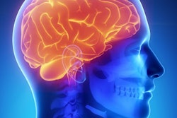
Radiologists must beware of normal brain images in cancer patients with neurological symptoms and adopt more sensitive techniques to catch brain metastasis. That's the view of Prof. Michael Forsting, who will deliver a keynote lecture on this topic today at the 100th German Radiology Congress (RöKo 2019), to be held from 29 May to 1 June in Leipzig.
The main danger in oncology patients is false-negative brain findings, particularly in patient subgroups, such as those with lung and breast cancers and renal cell carcinoma, for whom there is a high risk of brain metastasis. Further, there seems to be a real sense that brain imaging for metastasis is "easy" when, in fact, it isn't, he told AuntMinnieEurope.com ahead of the congress.
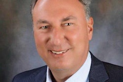
 Far from easy, brain metastasis detection requires expertise and care, says Prof. Michael Forsting.
"Radiologists need to be highly aware of any mismatch between imaging and clinical findings and change imaging strategy accordingly," noted Forsting, who is head of radiology at University Hospital Essen and one of three congress presidents for RöKo 2019. "The presence of brain metastasis and the extent of any metastasis will change therapy drastically."
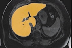
If normal gradient-echo sequences show no abnormalities when there are clinical symptoms, radiologists can take three routes. The first is to increase contrast dose, the second is to use new MR sequences such as black blood imaging that is more sensitive to intracranial and leptomeningeal metastasis, and third, they can use fluid-attenuated inversion-recovery (FLAIR) sequences in combination with contrast imaging.
These techniques are known but are not always at the forefront of radiologists' standard practice when imaging the brain, according to Forsting. He points to a tendency for radiologists of any level to trust in images rather than other evidence, despite the fact that some sequences are not sensitive enough. For example, MRI's limited spatial resolution makes micrometastasis difficult to spot.
In cancer patient subgroups, changing imaging strategy can increase the sensitivity of brain metastasis detection by 20%, he noted. In addition, increased sensitivity may reveal a higher number of brain metastases in a patient, affecting the therapeutic approach from stereotactic radiosurgery with a gamma knife, or surgical removal for one to three metastases, to whole-brain radiation for five or more metastases.
Forsting's presentation forms part of the international program, now a long-standing congress initiative, and is aimed at any radiologists involved in brain imaging to remind them of these existing yet relatively new possibilities for metastasis detection.
Emphasis on AI
At this year's centenary congress, artificial intelligence (AI) also will have a special place in the program.
"This is the first large congress in Germany that has chosen AI as a major theme. We want to discuss how AI will make our work better -- and easier," said Forsting, who expects the AI topics to attract a mixture of attendees from those who are fearful of the technology and those who are interested in unlocking its true potential.
Experts from radiology and general science will provide keynote lectures throughout the four-day congress. These lectures will be split into two categories: practical applications for AI to alleviate "boring" routine work, such as follow-up tumor imaging, plaque counting, and lung and breast cancer screening, and also radiomics, particularly for gleaning more information from images, such as molecular information from CT scans or tumor fingerprinting data, for example.
Forsting hopes that making AI a key theme will attract more attendees than last year, when attendance numbers slipped by 8% compared with 2017. He hopes the centenary congress will have over 7,000 attendees, compared with 6,145 in 2018 down from 6,673 in 2017.
 The poster for the 100th German Radiology Congress (RöKo 2019). Image courtesy of the DRG.
The 2018 figure simply reflected a broader trend of generally lower onsite registration across all medical congresses due to more widely accessible online information, he said. This year, however, there have been greater efforts to involve more technicians and industry figures -- the two groups that showed the biggest drop in attendance last year.
At RöKo 2018, technicians and radiography students totaled 1,312 (versus 1,469 in 2017), and industry delegates totaled 1,109 (1,389 in 2017). This represented a decline of 157 delegates (10.7%) and 280 attendees (20.2%), respectively.
To counter this trend, RöKo's organizers have approached about 15 AI-related smaller businesses, both startups and established companies, which will be showcasing their new products and solutions alongside the large vendors at the exhibition. Also, there has been a renewed drive to promote the AI congress theme to technicians, as the technology will have a significant effect on work such as MRI planning, according to Forsting.
"We're hoping that more of our radiology and other professional colleagues will join us this year," he added. "Germany continues to excel in radiology education, certification, and specialization, and we plan to continue sharing and learning both at a national and international level."