Dutch and Danish researchers have developed a fully automated technique to segment and collect measurements of the aorta from noncontrast 3D CT scans, according to an article published online on 23 January in European Radiology. The technique proved to be as effective as standard manual methods.
Measuring the aortic diameter on the 3D CT scans of patients with an aneurysm allows for the early detection of aortic dilatation and can ultimately help clinicians identify patients at risk of fatal conditions such as acute dissection, wrote lead author Zahra Gamechi, PhD, from Erasmus University Medical Center, and colleagues. Yet aortic aneurysms often go unmeasured because most patients are asymptomatic and do not undergo medical imaging.
To identify more of these high-risk patients, several groups have suggested that clinicians obtain aortic measurements of screening program participants who are already scheduled for a CT scan, the authors noted. This additional work, however, would be time-consuming and labor-intensive using traditional, manual measuring techniques.
Gamechi and colleagues thus developed a technique for automatically segmenting and measuring the thoracic aorta on noncontrast CT scans. Although several segmentation techniques exist that enable clinicians to collect aortic measurements automatically, they are mostly limited to use with contrast-enhanced imaging such as coronary CT angiography, according to the authors.
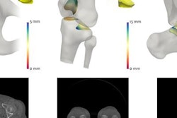
The researchers' automatic segmentation technique combines multiple image-processing steps, including a multiatlas registration algorithm, to localize and segment the aorta and major arteries on 3D CT scans. Then it calculates the average aortic diameter based on measurements from 13 cross-sectional slices taken at fixed intervals with respect to a single anatomical landmark.
The researchers validated their automated technique on the CT scans of 742 participants in the Danish Lung Cancer Screening Trial. Next, they compared the results with measurements of the same CT scans acquired using a standard manual protocol.
Overall, they found that their automatic segmentation method produced aortic measurements more quickly than manual segmentation and also with a similar degree of accuracy. The automated and manual segmentations overlapped extremely well, with a Dice similarity coefficient of 0.95. The mean surface distance between the manual and automatic segmentation surfaces also was very small at 0.56 mm.
For annotations, there was a minimal absolute error of 1.09 mm between manual and automatic diameter measurements, indicating a slight underestimation in size using the automated method. Nonetheless, the correlation between the diameter measurements was high: The intraclass correlation was 0.97 -- matching the level of agreement typically found among different manual measurements.
Collectively, these figures confirm that automatic segmentation of 3D CT scans can match the accuracy of manual segmentation -- opening wide the possibility of rapidly collecting aortic measurements from chest CT scans, the authors noted.
"The proposed automatic method is a promising technique to accurately and reproducibly assess subtle signs of aorta dilatation in nonelectrocardiogram-gated, noncontrast CT scans without any human interaction and could be used for efficient screening for aortic dilatation, as well as for monitoring of aortic change in clinical practice as part of a comprehensive CT analysis, including lung screening," they concluded.