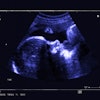
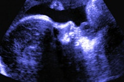
The use of ultrasound for pediatric appendicitis imaging has increased, but the modality remains underutilized in this area, according to research published on 22 August in the Journal of Pediatric Surgery.
A team led by Dr. Mark Slidell, from Johns Hopkins University in Baltimore, Maryland, U.S., found that CT remains the most used imaging method for diagnostic pediatric appendicitis in hospitals that don’t participate in the National Surgical Quality Improvement Program - Pediatrics (NSQIP-P).
“Adopting diagnostic strategies from NSQIP-P centers could optimize diagnostic imaging in children,” Slidell and colleagues wrote.
While ultrasound or MRI is recommended over CT for initial imaging when considering a diagnosis of appendicitis in children, CT is typically used first in adult patients with abdominal pain. The researchers highlighted that ultrasound and MRI reduce unnecessary radiation exposure and demonstrate high accuracy when imaging children.
The NSQIP-P aims to improve surgical care for pediatric patients. While hospitals that don’t participate in the program may provide care for children, they may not have specialized pediatric-focused initiatives and resources implemented, which can lead to imaging practices more appropriate for adult care.
Slidell and co-authors evaluated adherence to the “As Low As Reasonably Achievable” (ALARA) principle in U.S. hospitals over time. They included data collected between 2015 and 2021 for 115,186 patients under 18 years old undergoing appendectomy for acute appendicitis. The researchers compared rates of ultrasound, CT, and MRI usage between NSQIP-P hospitals and referring non-NSQIP-P hospitals.
Of the total study cohort, 66,303 (57.6%) were imaged in participating hospitals, 37,962 (33%) in nonparticipating hospitals, and 7,947 (6.9%) were imaged in both hospitals.
Ultrasound alone was used in 53.3% of the total hospitals, followed by CT alone in 25.1%, both ultrasound and CT in 16.4%, and MRI with or without CT or ultrasound in 2.6%. Also, non-NSIQP-P hospitals used less ultrasound than NSQIP-P centers (38.6% vs. 90.8%, p < 0.0001) and more CT (74% vs. 25.4%, p < 0.0001).
Finally, ultrasound’s overall use increased during the study period while CT’s use remained steady.
| Use of ultrasound, CT in diagnosing pediatric appendicitis | |||
|---|---|---|---|
| Modality and hospital type | 2015 | 2021 | p-value |
| Ultrasound | |||
| Overall | 68.5% | 72.3% | < 0.001 |
| NSQIP-P hospital | 71.7% | 70.9% | 0.28 |
| Non-NSQIP-P hospital | 18.8% | 25.7% | < 0.001 |
| CT | |||
| Overall | 43.1% | 43.2% | 0.07 |
| NSQIP-P hospital | 10.1% | 7.7% | < 0.001 |
| Non-NSQIP-P hospital | 71% | 59.8% | < 0.001 |
Despite the results favoring ultrasound’s use, the study authors expressed surprise that they didn’t observe “greater adherence” to the ALARA principles and efforts at reducing radiation exposure from CT scans in children.
“The identified plateau in CT rates may represent a need for further initiatives or alternative approaches to further reducing or eliminating CT usage in pediatric patients,” they wrote.
And while rapid-protocol MRI could help bridge gaps in imaging in this area, the authors noted that the modality’s growth in use for pediatric appendicitis imaging has been slow.
“Addressing these challenges will require more proactive measures,” they wrote. “The implementation of standardized in-hospital pathways has been shown to be an effective approach to improving adherence to ALARA principles in the process of diagnosing appendicitis.”
The full results can be found here.