PET/MRI scans have revealed positive findings for patients with late-life depression – namely that they have preserved “synaptic density" and thus are likely to respond to treatment, according to a group in Belgium.
Neuroscientists at the Leuven Brain Institute used hybrid brain imaging to explore connections between synaptic density (on PET) and gray-matter volume (on MRI) for the first time in a group of depressed patients, with a link between the two thought to be involved in neurodegenerative diseases.
“In contrast to Alzheimer’s disease, lower gray matter volume in late-life depression is not associated with synaptic density changes,” noted lead author Thomas Vande Casteele, MD, and colleagues. The study was published on 14 March in Translational Psychiatry.
Late-life depression has been consistently associated with lower gray-matter volume, the origin of which remains largely unexplained, the authors wrote. Recent PET findings in early-onset depression and Alzheimer’s disease suggest that synaptic deficits contribute to the pathophysiology of these disorders and therefore contribute to lower gray-matter volume, they suggested.
However, the hypothesis linking synaptic density with depression has only been investigated in animal stress studies and postmortem depression studies focusing on the hippocampus and prefrontal cortex, the authors added.
Thus, in this study, the researchers performed a prospective analysis to measure synaptic density and gray-matter volume in a group of patients. For the PET imaging, they used a radiotracer named carbon-11 (C-11) UCB-J, which binds to a glycoprotein expressed in virtually all synapses.
The group enrolled 24 currently depressed adults with late-life depression (average age, 73 years old) and 36 age- and gender-matched healthy controls (average age, 70 years old). Participants underwent simultaneous C-11 UCB-J PET and 3D T1- and T2-weighted fluid-attenuated inversion recovery (FLAIR) MRI on a 3-tesla PET/MRI scanner.
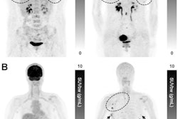
 PET/MRI images used in the analysis: Axial (left, through the prefrontal volume-of-interest) and coronal (right, through the hippocampal volume-of-interest) overlay of a partial volume corrected carbon-11 UCB-J PET image (right subparts) registered to the anatomical T1-weighted MRI background for delineation.Image courtesy of Translational Psychiatry
According to the findings, the late-life depression group showed a trend in lower gray matter volumes in the hippocampus (p = 0.04), mesial temporal (p = 0.02), and prefrontal cortex (p = 0.02) compared to the healthy control group. However, no group differences in C-11 UCB-J binding were found in these regions, nor were there any associations between C-11 UCB-J and depressive symptoms, they wrote.
“Our finding suggests that there is no regional difference in synaptic density as measured by [PET] C-11 UCB-J across several cortical regions implicated in late-life depression,” the group wrote.
As this was the first prospective study of its type, additional research is required to confirm the research, the authors noted. However, from a therapeutic standpoint, they noted that preserved synaptic density in late-life depression may be an “encouraging finding.”
“Preserved synaptic density may suggest that the aging brain in [late-life depression] retains its capacity for neuroplasticity and remains amenable to therapeutic interventions,” the researchers concluded.
The full study is available here.