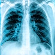
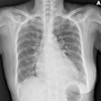
Dynamic chest radiography (DCR) shows potential as a tool to investigate lung health in people with cystic fibrosis (CF), according to research published on 13 February in Clinical Radiology.
This study is the first to describe diaphragm motion and projected lung areas using DCR in adults with stable disease, noted lead author Dr. Thomas FitzMaurice, of the University of Liverpool in the U.K., and colleagues.
“We have demonstrated DCR to be a versatile imaging technology that produces measurements of thoracic motion that have significant associations with spirometry,” the group wrote.
The lifespan of people with CF has improved significantly in recent years and is likely to rise further with the increasing availability of new drugs to treat the disease, the authors explained. Spirometry, specifically forced expiratory volume of air in 1 second (FEV1), is a prognostic marker in cystic fibrosis, and alongside chest x-ray findings is the primary method for assessing lung health in patients. Yet FEV1 may not always reflect the severity of the airway obstruction, they noted.
Alternatively, DCR, also referred to as dynamic digital radiography, is a low ionizing radiation x-ray system that produces wide field-of-view fluoroscopic-style images of the thorax in motion. Previous studies have shown that DCR can measure diaphragm motion in healthy individuals, people with chronic obstructive pulmonary disease, and interstitial lung disease.
To further explore the use of the technology, in this prospective study, the group tested whether it can monitor subtle changes in pulmonary physiology in adults receiving treatment for cystic fibrosis.
The researchers assessed 129 participants who underwent DCR and spirometry during their annual reviews between December 2019 and October 2020 instead of standard chest x-rays usually performed. DCR images were acquired with patients in the erect position over 10 seconds, at 15 frames per second.
Participants were instructed by a respiratory physician or radiographer to take a tidal breath, followed by a deep breath to full inspiration, followed by a passive breath out to end-expiration. In addition, spirometry was performed, and the researchers recorded FEV1, forced vital capacity (FVC), and FEV1/FVC ratios.
According to the analysis, several DCR parameters correlated with spirometric parameters, with the strongest correlation seen between changes in patients’ projected lung area (PLA) and FEV1. Patients’ average inspiratory lung area on DCR also correlated with FVC and with FEV1/FVC ratios, the group reported.
“This study is the first to describe diaphragm motion and projected lung areas using DCR in nonexacerbating adult [people with cystic fibrosis],” the authors wrote.
Future studies could include comparisons between DCR with similar low-dose imaging techniques, such as ultralow dose CT, as well as work to further validate and standardize the use of DCR, the group noted.
“The features of low ionizing radiation dose, ease of setup, and the simultaneous acquisition of diagnostic chest imaging may make DCR an attractive modality to monitor lung health of [people with cystic fibrosis],” the group concluded.
The full study is available here.


