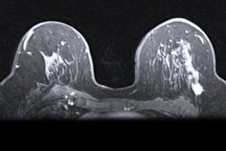
Radiologists need to remember women undergoing breast MRI often have metallic biopsy clips placed within or adjacent to a lesion, producing artifacts that can lead to misinterpretation, Austrian investigators have emphasized in new research.
"When analyzing diffusion-weighted images (DWI) and apparent diffusion coefficient (ADC) maps, one should always be aware of the presence of clips in the patient in question," Christian Kremser, PhD, a medical physicist in the Department of Radiology at Medical University of Innsbruck, told AuntMinnieEurope.com.
"Signal alterations produced by these biopsy marker clips could render the evaluation of the tissue near the marker impossible or lead to misinterpretations," he added, noting that the size of the lesion must be taken into account to ensure that in subsequent MR exams the tissue of interest can be properly assessed with DWI.
Kremser and his clinical colleagues at Innsbruck decided to study the artifacts produced by four different clips on DWI obtained with a readout-segmented sequence (REadout Segmentation Of Long Variable Echo trains, RESOLVE) and their influence on ADC maps. They presented their results in a poster at the annual meeting of the International Society for Magnetic Resonance in Medicine (ISMRM) in May.
 Size of artifact at 1.5 tesla for a range of investigated biopsy markers. All tables and figures courtesy of Christian Kremser, PhD; Dr. Birgit Amort; Dr. Martin Daniaux; and the ISMRM.
 Size of artifact at 3 tesla for biopsy markers.
All scans were performed on two 1.5-tesla and 3-tesla whole-body MR scanners (Magnetom AvantoFit and Magnetom Skyra, Siemens Healthineers) using an 18-channel breast coil. For each clip, a cylindrical agar phantom was prepared, and the breast coil was loaded with two of these phantoms at the left and right side. After an empty measurement, one clip per phantom was inserted at a depth of 6 cm by means of the biopsy needles provided with the clips.
The DWI images and ADC maps were evaluated using the hospital PACS and Impax EE software (R20 XVII SU1, Agfa). The size of the artifacts was larger and more prominent on DWI images with b=800s/mm2 as compared with ADC maps. The Bard UltraClip Dual Trigger showed the largest artifact, followed by the Tumark Vision and Tumark Professional clip. The smallest artifact was observed for HydroMark Breast Biopsy Site Marker.
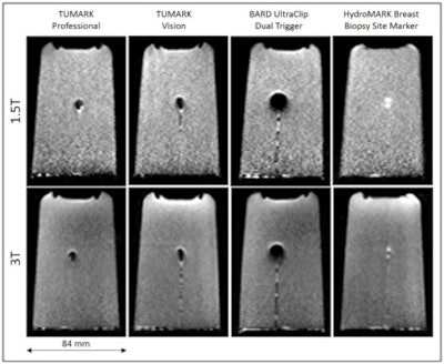 Maximum extent of the obtained artifacts for DWI images (b=800 s/mm2)
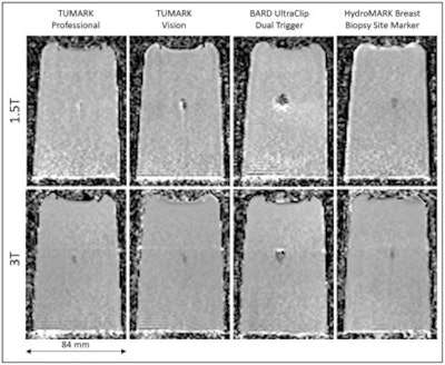 Maximum extent of the obtained artifacts for ADC maps.
Whereas most clips showed a clear signal void on the DWI images, the HydroMark device presented with a marked signal increase, both at 1.5 tesla and 3 tesla. On ADC maps, the observed ADC changes (increased or decreased ADC values) due to the artifact varied between clips and field strength.
Overall, the researchers found that artifacts vary in shape and size between different clips, DWI images and ADC-maps. For the marker with the smallest artifact, the minimum size of detectable lesion seems to be approximately 10 mm.
Need for caution
ADC values in regions where visible signal alterations on DWI images (b=800s/mm2) due to the clip are seen should be interpreted with caution. Even for the clips with the smallest artifact the size of these regions can measure 10 mm to 12 mm.
"The fact that stainless steel clips produced the largest artefact compared to nitinol and titanium was broadly in line with our expectations," said Kremser, who has worked on MRI projects at Innsbruck since 1993. "However, it was surprising that on ADC maps the observed artefacts were slightly smaller than on the diffusion weighted images. In any case it was a surprise that there has been no publication on this yet."
The team now plans to extend their studies to the whole recommended MR protocol for breast MRI and also look into echo-planar diffusion weighted imaging sequences.


