Award-winning research shows a well-crafted audit can substantially raise awareness of vasa previa and lead to a more rapid and accurate diagnosis of this complication of pregnancy.
"Vasa previa is a rare but potentially catastrophic cause of antepartum hemorrhage and can result in fetal death. When diagnosed prenatally, it can make a major impact on the management of these cases," explained Dr. Sandeep Tiwari, a consultant radiologist at Nepean Radiology in Sydney, Australia.
Vasa previa is defined as aberrant fetal blood vessels crossing over or within 2 cm of the internal cervical os. These blood vessels are unprotected by the umbilical cord or placenta and run through the amniotic membranes, according to Tiwari and his colleagues, whose e-poster presentation won a top award at the recent Royal Australian & New Zealand College of Radiology (RANZCR) annual scientific meeting.

There are two types of vasa previa: Type 1 occurs when abnormal fetal vessels connect a velamentous cord insertion to the main body of the placenta; type 2 occurs when fetal vessels connect a bilobed placenta or a placenta with succenturiate lobe.
Guidelines for sonographers
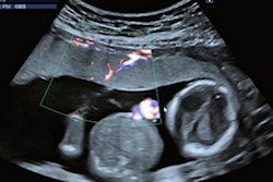
The Australasian Sonographers' Association (ASA) released guidelines in May 2020 on vasa previa diagnosis in midtrimester ultrasound. ASA emphasizes that a dynamic sweep of the transducer with color Doppler over the lower uterine segment should be performed and documented at every mid-trimester ultrasound scan.
Tiwari and his colleagues performed an audit to assess their practice in this area. The aim was to raise awareness among the sonographers about the new guidelines and improve our efficacy in the diagnosis of vasa previa.
"Compliance/performance rate of color box assessment over the internal os by sonographers in mid-trimester ultrasound was assessed. In our initial audit the compliance rate was 50% in March 2020, which was improved to 88% in the re-audit in March 2021," noted Tiwari, who previously worked as a radiologist at Nottingham University Hospital, U.K.
The plan is to continue to improve the department's efficacy in assessment of vasa previa by regular discussion with sonographers and performing further reaudits, he concluded.
Editor's note: You can view the authors' full poster on the RANZCR section of the Electronic Presentation Online System (EPOS) on the European Society of Radiology's website.


