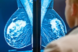
Digital mammography and digital breast tomosynthesis (DBT) inadvertently performed during pregnancy do not pose a significant radiation risk for pregnant women or their fetuses, according to Portuguese research published on 24 September in Radiography.
A team led by Dr. Ana Raquel Cepeda Martins from Igamaot in Lisbon found that the absorbed radiation dose values during mammography to the abdomen are negligible, hinting at a negligible dose to the uterus, even in cases of overradiation.
"The findings in this study could contribute to reducing the psychological burden of pregnant women undergoing breast screening examinations," Martins and colleagues wrote.
Mammography and DBT have been touted for their success in detecting breast cancers. However, both methods deliver a radiation dose between 2 and 4 mGy, depending on breast thickness and glandularity, the researchers wrote.
Pregnant women are usually discouraged from undergoing medical imaging with imaging modalities that use ionizing radiation. But some exams do occur, such as among women who discover they are pregnant only after an exam. In any event, the literature is scarce for fetal dosimetry related to mammography.
Therefore, the team wanted to estimate the level of radiation dose to the uterus in standard mammography and DBT. They used an anthropomorphic phantom with plates simulating breasts with a thickness of 6 cm. Craniocaudal and mediolateral oblique views were used for mammography and DBT examinations.
They worked in two main scenarios, each of which could result in different levels of radiation dose. The first one saw the exposure parameters preselected directly by the imaging system, while in the second scenario saw the maximum exposure parameters chosen.
In the first scenario, dosimetry measurements did not detect significant dose values in the abdomen. Mean glandular doses meanwhile were found to be in close agreement with data available in previous studies, between 1.5 and 1.9 mGy.
In the second scenario, there was no significant difference in mean glandular dose estimation between the different views, with abdominal air kerma values for DBT being 0.049 mGy (craniocaudal) and 0.004 mGy (mediolateral oblique) respectively. Craniocaudal abdominal views with mammography meanwhile saw an air kerma value of 0.026 mGy, with no significant detected value in mediolateral oblique view.
The researchers noted that an average fetal dose less than 50 mGy has not been associated with any significant adverse fetal effects.
"It is safe to say that mammography and DBT could be considered safe, posing no significant risks, during pregnancy," they wrote.
However, the study authors also noted that each case should be evaluated according to patient history and that when no consensus is reached, the linear no-threshold model could be "always considered" to keep the radiation dose to the fetus as low as reasonably achievable.
Martins and colleagues said having accurate knowledge of radiation doses in mammography and DBT will help raise awareness among breast imagers, as well as improve radiation risk communication skills and help reduce anxiety in pregnant women.


