Evaluation of longitudinal strain by CT is fast emerging as a useful tool to stratify low-risk patients with pulmonary embolism (PE), according to new research being presented at this week's European Society of Cardiology (ESC) annual congress.
Right ventricle dysfunction remains the leading cause of mortality in intermediate and high-risk PE, explained Dr. Jorge Armando Joya and colleagues from the Instituto de Cardiología y Medicina Vascular, TEC Salud and Centro de Investigacion Biomedica del Hospital Zambrano Hellion, Escuela de Medicina, Tecnologico de Monterrey, Mexico.
"Current ESC guidelines recommend a multimodal stratification, including echocardiographic or CT high-sensitivity cardiac troponin for intermediate and high-risk PE. Inherently, the right ventricle has an overly complex anatomy with a trabeculated structure, which makes its imaging evaluation difficult," they noted in an ESC 2021 e-poster.
Study objectives
Longitudinal strain is a useful technique that quantifies the myocardial deformation without geometric assumptions, allowing the assessment of the right ventricle myocardial function, the authors continued. Its validation as a marker of mortality has been tested in several cardiovascular diseases, but the CT strain rate for right ventricular dysfunction in the context of PE has not been standardized.
Therefore, Joya and colleagues sought to detect early right ventricle abnormalities in patients with acute PE. They determined the longitudinal strain value by vectorial epicardial pixels movement, or tissue tracking, using cardiac CT. They also wanted to discover if there is a significant difference between right ventricle longitudinal strain in patients with acute (low, intermediate, and high risk) PE compared with healthy controls.
The team conducted an observational retrospective single-center study involving patients admitted to hospital with PE proved by CT angiography between 2016 and 2020. To be included, patients had to sign an informed consent form, be 18 years old or over, and have a diagnosis of acute PE after a multiphase CT angiogram. The control group comprised patients who had had a check-up and noncardiac chest pain causes with CT angiography negate for PE and no evidence of valvular disease and a coronary calcium score of 0 Agatston units.
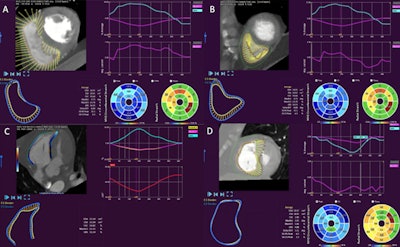
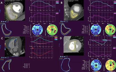
"We evaluated the right ventricle in the four-chamber views from the multiphase CT angiogram, which were acquired every 10% of the cardiac cycle with a small field view," the researchers noted. "Posteriorly, the images were reconstructed to assess the ventricle dynamics and obtain the longitudinal strain value using tissue tracking CT software."


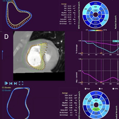
The Mexican group evaluated the right ventricle four-chamber images from the multiphase CT angiogram. The images were reconstructed to assess the dynamics and the longitudinal strain by tissue-tracking CT software. (A) No right ventricle pathology (diastole). (B) No right ventricle pathology (systole). (C) McConnell's sign by CT strain. (D) Impaired right ventricle function due to pulmonary embolism. Image courtesy of Dr. Jaime Alberto Guajardo-Lozano, Dr. Jorge Joya-Harrison, and colleagues.
An experienced cardiologist then evaluated the result, after which a receiver-operating curve (ROC) was created to assess the longitudinal strain performance for right ventricle dysfunction detection. The investigators calculated the Youden index for every value to identify the best cut point, considering the area under the curve, sensitivity, and specificity. They considered a p value of < 0.05 for statistical significance with a confidence interval of 95%.
Key findings
A total of 53 patients with a diagnosis of PE were admitted to the hospital, of whom only 10 fulfilled all the inclusion criteria. The control group consisted of 500 patients, of whom only 33 had a complete multiphase sequence and a negative result for PE or other cardiac diseases. In the PE group, the gender mix was 70 to 30, men to women, compared with a ratio of 58 to 42 in the control group.
There was an overt tendency for alteration of the right ventricle longitudinal strain in patients with PE compared with the control group (-8.41% vs. -21.09%, p < 0.001), according to Joya and colleagues. The area under the curve for the cut-point of -18.25% for the longitudinal strain was 0.89, with a sensitivity of 91% and specificity of 90% for detecting right ventricle dysfunction.
The lower right ventricular fractional shortening percentages also correlated with PE diagnosis versus controls (12.3 vs. 31 respectively). Another expected finding was a decreased oxygen saturation in PE patients vs. the control group (92.1 ± 5.7 vs. 97 ± 2.1, p > 0.031), the authors stated.
A major limitation was the size of the population, they admitted. "We kept only 18.86% of patients in the initial universe due to incomplete acquisition of multiphase sequence in the CT angiogram. There is no reference value of right ventricle longitudinal strain in the general population."
Overall, they propose the cut-point value of -18.25% for longitudinal strain by cardiac CT to detect early right ventricular dysfunction in patients with acute PE, and a value of -21.09% cutoff point for healthy patients.
Correlation and clinical impact have yet to be studied, and further research involving a larger population size is needed to confirm these results, they concluded.
"Further studies involving CT strain are in progress as to evaluate not only global right ventricular longitudinal strain, but also circumferential and free wall strain," cardiology fellow Dr. Jaime Alberto Guajardo-Lozano told AuntMinnieEurope.com. "Right atrial strain measurements are also an area of interest in patients with pulmonary hypertension and will be evaluated as a prognostic marker."
Editor's note: "ESC Congress 2021 - The Digital Experience" runs from 27 to 30 August. Some material is already available on the congress website.


