The global MRI community has lost one of its major pioneers in Ian Robert Young, PhD, the well-known medical physicist who died on 27 September. Modern clinical brain and body imaging use many pulse sequences and other techniques first demonstrated by him and his group, and he was responsible for a lot of the early work in MRI of multiple sclerosis (MS).
Ian was born in London on 11 January 1932, the eldest of three sons. His father, John Stirling Young, was Regius Professor of Pathology at the University of Aberdeen from 1937 to 1962. Ian was educated at Sedbergh School in Cumbria, northwest England, and studied natural philosophy at the University of Aberdeen. He completed his doctorate there under the supervision of Reginald V Jones (1911-1997), who was famous for many feats of scientific intelligence and the development of countermeasures during World War II.
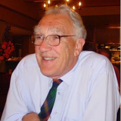
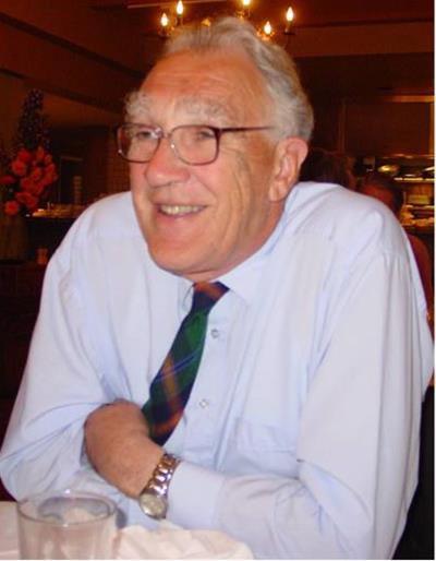
 Ian Robert Young, PhD.
After working for Hilger and Watts, Evershed and Vignoles, and Evershed Power Optics, Ian joined the Electrical and Musical Industries (EMI) Central Research Laboratory in August 1976. EMI was established in 1931 as a vertically integrated company producing sound recordings as well as recording and playback equipment. To this was added TV broadcasting and stereo sound as well as radar during World War II under the direction of Alan Blumlein.
After the war, the company specialized in entertainment and defense until quite unexpectedly, and at very little cost to the company, Godfrey Hounsfield developed the world's first head CT scanner. This transformed neuroradiology. In 1975 he followed this with the CT5000 body scanner. It was a major departure for the company that had no previous track record in x-ray technology or medical equipment, but following the invention of MRI by Paul Lauterbur in 1973, EMI decided to expand its role in medical imaging and established a nuclear magnetic resonance (NMR) group.
The NMR group began with a Walker 0.1-tesla resistive magnet and, under the direction of Hugh Clow and Ian Young, produced the world's first published human MR image of the brain in November 1978. This was followed by an inversion recovery (IR) image of Ian Young's brain in autumn 1979 that showed very high gray-white matter contrast. The image was shown by Godfrey Hounsfield in his Nobel prize lecture on computed medical imaging on 8 December 1979, together with his explanation of the image contrast in terms of differences in T1 (see figures below).
 Above: Godfrey Hounsfield's diagram relating signal amplitude to the log of tissue T1 including gray brain and white brain. Hounsfield presented this during his Nobel lecture on 8 December, 1979. The curve opposite the numeral 2 shows white matter with higher amplitude than gray matter as seen on the IR image (TR/TE = 1000/300 ms) of Ian Young's brain. Below: Young's brain obtained at the EMI Central Research Laboratory in autumn 1979.
Ian Young added to the gradient echo and spin echo (SE) sequences in 1980. With funding from the scientific and technical services branch of the Department of Health and Social Security headed by Gordon Higson (who had worked so fruitfully with Godfrey Hounsfield), his group built a 0.15-tesla system based on the world's first commercial large-bore cryomagnet. This was built by Oxford Instruments under the direction of Martin Wood. The MR system was shifted to Hammersmith Hospital in London and patient imaging began in March 1981.
There was a tremendous sense of excitement in the department at that time as Graeme Bydder brought new MR images to the lunchtime meetings, fascinating the junior radiologists. In November 1981, Ian published a study of 10 MS patients, in which 112 lesions were detected with MRI and only 19 were seen with CT. It was a quantifiable, decisive, clinical advantage for MRI over CT, and lead to a flurry of activity. Oxford Instruments' order book for magnets increased from 1 million pounds (1.12 million euros) in 1981, to 25 million pounds (28 million euros) in 1982.
The success with the IR sequence was followed by David Bailes' development of the heavily T2-weighted SE sequence used in a study of 32 brain patients that was published in July 1982, as well as for studies of the posterior fossa where CT was degraded by beam hardening, and MR had a significant clinical advantage.
RSNA 1982: The big breakthrough
The world of MRI was turned on its head at the November 1982 meeting of the RSNA in Chicago, when GE showed very high quality brain images obtained at 1.5-tesla -- 10 times the field strength that Ian and many others were operating at. The GE system was aggressively marketed even though the company had no product to sell. Customers were strongly advised not to buy other companies' lower field systems and to buy the 1.5-tesla GE product when it became available.
This was the beginning of the "field wars" that dominated MRI for the next four to five years. Ian and his group developed new receiver coils, used low-bandwidth acquisitions, the short inversion time (STIR) pulse sequences, heavily T2-weighted gradient echo sequences, and motion artifact control techniques (e.g., respiratory ordered phase encoding, ROPE). They also used the contrast agent gadolinium-DTPA developed by Hanns-Joachim Weinmann of Schering and showed it was not a cut and dried issue; it was possible to perform clinically competitive studies at low field using much cheaper systems.
Jo Hajnal of Ian's group later developed the fluid-attenuated inversion-recovery (FLAIR) sequence and another member, David Gilderdale, produced internal prostate, rectal, cervical, and vaginal coils. David Bryant and others of the group built a 1.5-tesla spectroscopy system and published over 200 papers on this subject with basic science and clinical groups. Alasdair Hall also built the first dedicated neonatal system in 1995.
MS advances
MS remains the disease par excellence associated with MRI. James Prichard, neurologist at Yale, said, "Ian's early paper on MRI detection of clinically silent demyelinating lesions in MS was the earliest major, and still one of the most important, MRI discoveries pertinent to neurology. It showed that what we neurologists had always considered an unpredictable intermittent disease is actually a continuously progressive pathological condition of which only the clinical expression is intermittent. That insight both deepened our understanding of the basic processes at work and provided a new treatment evaluation metric that greatly sped up treatment research."
Ian's work was widely recognized. He was awarded an Order of the British Empire (OBE) award in 1986 and was elected a Fellow of the Royal Academy of Engineering (FREng) in 1988 and a Fellow of the Royal Society (FRS) in 1989. He was also elected president of the International Society for Magnetic Resonance in Medicine (ISMRM) in 1991-1992, and received the society's gold and silver medals, as well as medals from the Royal Academy of Engineering, the Institute of Physics, and the Royal Society. He is survived by his wife Sylvia, their children Graham, Neil, and Fiona, and their six grandchildren.
Ian's final project was developing, with Graeme Bydder and Martyn Paley, an online history of MRI and spectroscopy in the U.K. This is a wonderful resource and can be found at https://mrishistory.org.uk.
Graeme Bydder is director and professor in the department of radiology at the University of California, San Diego (UCSD). Dr. Adrian Thomas is secretary of the International Society for the History of Radiology and honorary historian at the British Institute of Radiology (BIR).
The comments and observations expressed herein do not necessarily reflect the opinions of AuntMinnieEurope.com, nor should they be construed as an endorsement or admonishment of any particular vendor, analyst, industry consultant, or consulting group.


