PET/MRI was proposed first in the 1990s by Paul Marsden, PhD; Simon Cherry, PhD; and others to invigorate in vivo small animal imaging. The first records of champagne date back to 1531, when Benedictine monks near Carcassone proposed a sparkling wine called Blanquette de Limoux. With a time difference of 500 years between the origins of champagne and integrated PET/MRI, why would one associate them together?
PET/MRI and champagne have a lot in common. On the face of it, both are expensive and even exclusive, befitting the "rich and famous."1 But "expensiveness" lies in the eyes of the beholder. Imaging examinations, particularly hybrid imaging exams, frequently are regarded as expensive and their cost-effectiveness questioned, even by regular users of such hybrid modalities.
This is extraordinary, given what hybrid imaging can offer, including accurate and timely diagnosis, increased patient comfort and better experience compared with standalone imaging, and the potential to facilitate personalized treatment. In fact, to date, less than 1% of cancer management costs are incurred by imaging.
Likewise, a bottle of champagne is expensive only when the consumer does not value it. It takes experience and attention to produce a good product, and champagne is best when the vineyards have been established over years, when the weather conditions have been ideal, and when the grapes from a particular harvest have been handled with care by trained winemakers. The same holds true for PET/MRI. Much like the Benedictine monks have evolved their sparkling wine production over many years, it has also taken a long time for the concept of an integrated PET/MRI system to reach subsequent production.
Champagne improves with age. In fact, for good champagne to be consumed, there should be several years between the harvest and consumption. This is because champagne ages well in the bottle under the right conditions. Again, the similarities with PET/MRI are striking. When a fully integrated PET/MRI system was introduced for the first time in 2006, there was hype and enthusiasm, and perhaps inflated expectation. In analogy to the periodic turning of bottles of champagne, it has taken numerous turns from the PET/MRI community -- including engineers, physicists, technologists, and medical experts -- before this imaging modality has matured.2
But they have succeeded. Those who can afford an integrated PET/MRI can indulge, if you permit the use of this paraphrase, in an optimized imaging system that provides the user with quantitative and complementary "anato-structural-metabolic" image information. Like champagne, so will PET/MRI mature. Already, clinical applications have evolved beyond the simple image fusion of PET- and MR-based image information.


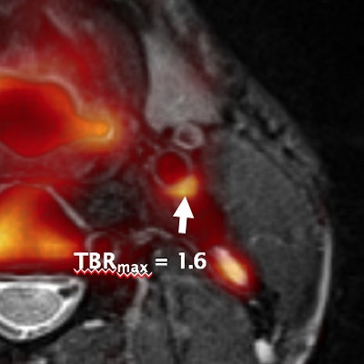
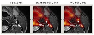
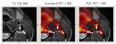
Figure 1: Integrated PET/MRI using [Ga-68]-Pentioxafor (CXCR4) for imaging vulnerable plaque. Correlated MR-based anatomical information can be used for partial volume correction (PVC) of PET tracer uptake in small regions, such as plaques. In this case, T2-weighted turbo spin-echo (TSE) MR image reveals a relatively stable plaque, with regular borders and uniform morphology. The standard PET image does not show increased CXCR4 uptake, while the PVC PET image shows a slight increase of CXCR4 uptake; the tumor-to-background ratio (TBR) increases from 1.3 to 1.6. Images courtesy of Medical University Vienna.
Novel concepts toward intrinsic compensation of involuntary patient motion are being worked on, a prerequisite for subsequent partial volume correction (figure 1), or anatomic-guided image reconstruction. The better the alignment of the two sets of imaging information, the more valuable the clinical information of an integrated PET/MRI examination (figure 2). Also, the palette of PET/MRI scenarios is being expanded through the adoption of the concept of an automated image-derived input function as a prerequisite for parametric imaging, as well as through the adoption of novel radiotracers, such as gallium-68 prostate-specific membrane antigen (Ga-68 PSMA) for multiparametric PET/MR imaging of prostate cancer.


Figure 2: Combined F-18 FDG PET/MRI of a primary upper rectal cancer demonstrates a metabolically active tumor with restricted diffusion and type II enhancement curve. Images courtesy of King's College London.
It is good to enjoy a wonderful bottle of champagne. In fact, many people go back time and again to retaste and re-experience the same ambiance in which a bottle was enjoyed. Undoubtedly, "retaste" is a potential downside of PET/MRI today.
In contrast to PET imaging alone, few studies exist today that demonstrate a sufficient reproducibility and repeatability of PET/MR information. Such evidence is, however, needed if we wish to leverage larger cohort clinical insights into PET/MRI that will be required in due course by the insurance providers. We need to be prepared for that through the engagement of all experts involved, across countries, disciplines, and associations; a testimony to such efforts is the recently awarded Horizon 2020 Innovative Training Network (ITN) called HYBRID (Healthcare Yearns Bright Researchers for Imaging Data), which resembles a consortium of multiple European academic, commercial, and educational partners. The project is due to start on 1 November 2017.
If we manage this process, we will be able to enjoy PET/MRI as an asset of personalized patient management, and, thus, indulge in a perfectly chilled and mouthwatering glass of champagne. We should drink to that.
Thomas Beyer, PhD, is a professor of physics of medical imaging at the Center for Medical Physics and Biomedical Engineering, QIMP Group, at Medical University of Vienna. Dr. Vicky Goh is a professor and the chair of clinical cancer imaging and the deputy head in the department of cancer imaging in the Division of Imaging Sciences and Biomedical Engineering at King's College London.
References
- Beyer T, Hacker M, Goh V. PET/MRI: Knocking on the doors of the rich and famous. Br J Radiol. 2017;90(1077):20170347.
- Bailey DL, Pichler BJ, Gückel B, et al. Combined PET/MRI: From Status Quo to Status Go. Summary Report of the Fifth International Workshop on PET/MR Imaging; February 15-19, 2016; Tübingen, Germany. Mol Imag Biol. 2016;18(5):637-650.
The comments and observations expressed herein do not necessarily reflect the opinions of AuntMinnieEurope.com, nor should they be construed as an endorsement or admonishment of any particular vendor, analyst, industry consultant, or consulting group.


