German researchers have developed a low-cost 3D printing technology that can be used to create patient-specific organ models of varying sizes and shapes, according to a study published in the December issue of the Journal of Nuclear Medicine.
The process for clinical prototyping has produced a 3D kidney phantom for quantitative SPECT/CT imaging to help optimize radiation dose for radionuclide therapies for each individual patient (JNM, Vol. 57:12, pp. 1998-2005).
Using the inexpensive models for direct calibration is particularly important for imaging systems that are prone to poor spatial resolution and ill-defined quantification, such as SPECT/CT, said lead author Johannes Tran-Gia, PhD, from the University of Würzburg, in a statement from the Society of Nuclear Medicine and Molecular Imaging (SNMMI).
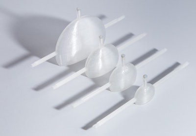 The manufactured set of kidney phantoms can be used for patients of different sizes and ages: newborn, 1-year-old, 5-year-old, and adult. All images courtesy of the University of Würzburg.
A set of four single-compartment kidney dosimetry phantoms can be filled with volumes ranging from approximately 8 mL, representing newborn patients, to 123 mL, for adults. The phantoms are based on outer kidney dimensions provided by Medical Internal Radiation Dose (MIRD) guidelines.
Based on these designs, refillable, waterproof, and chemically stable models were manufactured with a fused deposition modeling 3D printer. Nuclide-dependent SPECT/CT calibration factors for technetium-99m, lutetium-177, and iodine-131 were then determined to assess the accuracy of quantitative imaging for internal renal dosimetry.
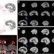
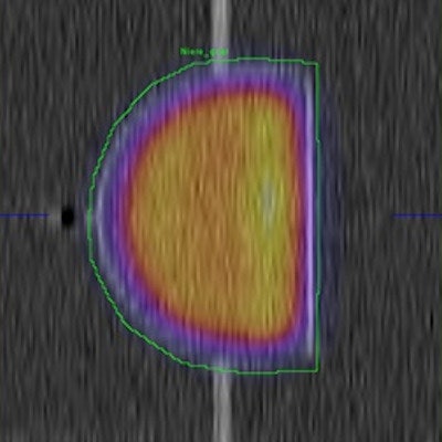
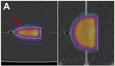 SPECT/CT reconstructions and volumes of interest were used to determine calibration factors for the adult kidney filled with lutetium-177 (A) and the corresponding sphere filled with iodine-131 (B).
Although the kidneys were modeled with a relatively simple one-compartment design, "the study represents an important step toward a reliable determination of absorbed doses and, therefore, an individualized patient dosimetry of other critical organs in addition to kidneys," Tran-Gia said.

