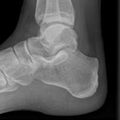
The severity of a soft-tissue ankle injury as determined by initial classification based on radiological findings is not always an accurate predictor of outcome at six months, according to new research from the U.K.
A prospective observational cohort study involving 100 patients from the John Radcliffe Hospital's accident and emergency (A&E) department in Oxford was conducted between February 2013 and August 2015. Patients were included if they were over 18 years old with a soft-tissue injury of the ankle not requiring radiographs, or where plain radiographs were deemed normal.
At the initial examination, within five days of injury, patients underwent a low-dose extremity CT scan, an ultrasound scan, and a disability assessment, explained lead author Dr. David J. Wilson, a consultant musculoskeletal (MSK) radiologist at St. Luke's Radiology, in an e-poster presentation at ECR 2016. St. Luke's Radiology provides specialist MSK clinical and imaging services and is based in Headington, Oxford.
Any abnormalities identified that warranted further investigation were referred back to A&E or the trauma and orthopedics service at the John Radcliffe Hospital. Of these, any patients with fractures or injuries that required orthopedic management were subsequently excluded from the follow-up phase of the study, he added. Follow-up was conducted at three and six months postinjury, at which time an ultrasound scan was repeated by a consultant radiologist and a physical examination was performed by a physiotherapist.
All 100 recruited patients attended the initial examination, with a cohort mean age of 33 (18 to 68; 53 male, 47 female). The number of ankle injuries included in the study totaled 101 because one patient injured both ankles. Following the initial examination, five patients were excluded from follow-up. After initial CT and ultrasound scans, 68 patients were deemed to have simple soft-tissue ankle injuries and 28 had complex. In the simple group, loss to follow-up was 47% at three months and 60% at six months. For the complex group, loss to follow-up was 46% at three months and 36% at six months, wrote Wilson, who is also president of the British Institute of Radiology (BIR).
Types of injury
The most common injury was to the anterior talofibular ligament at 83%, with complete tears found in 67% of injuries. Deltoid ligament injuries were the second most common at 39%, with only 3% complete tears and the vast majority being sprains. The calcaneofibular ligament and talonavicular ligaments were also common injuries amongst the cohort, and 15 complete tears of the anterior inferior tibiofibular ligament were identified, of which none showed diastasis on dynamic imaging.
"It was very common for people to sustain multiple soft-tissue injuries with 31% injuring a single anatomical tissue, 33% two, 20% three, 8% four, and 4% five," Wilson stated.
The visual analogue scale (VAS) pain scores and quality of life (QoL) questionnaires showed overall improvement throughout the course of the study. There was minimal difference between both groups at all stages for mean VAS scores, with very similar ranges. In the QoL score, there was again minimal difference between the group means, particularly at the initial assessment and at the six-month point, but there was a slight difference at three months, with a much wider range of scores in the complex group, the authors found.
"One may have assumed that a more serious injury leads to worse outcomes and a delayed recovery compared to simple injuries. Although there were some small differences, this was not demonstrated in the data collected and analyzed in this study," they noted. "From the cohort observed, it would appear that on average patients recover at a similar rate. That is not to say that some people did not take longer than others and experience more pain or disability."
This study also emphasized the importance of the management of swelling for optimal functional recovery, which patients may not be addressing ideally, continued Wilson et al. Recovery for the lay person following an ankle injury may require improved advice and education to optimize their return to full function.
Study limitations
Many elements that might have affected a patient's recovery were not considered in this study, they admitted. Age, method of injury, compliance with PRICE (protection, rest, ice, compression, elevation) advice, allied health professional treatment, activity levels, and exercises performed are some of the main limitations.
Ankle injuries are one of the most common presentations to emergency departments, and there are an estimated 302,000 new cases per year in the U.K. alone. The outcome of these patients is often unknown and although many go on to a full recovery, up to 30% can develop longer-term dysfunction and disability, according to Wilson.
"The initial management of ankle injuries varies by department and clinician, which is often dictated by the severity and presentation of the patient," he pointed out. "Most commonly, the investigation of choice in the emergency department is plain radiographs using the Ottowa Foot and Ankle Rules to determine suitability."
When an injury appears severe or abnormalities are noted on radiographs, the patient is routinely referred to fracture clinic for further assessment and management, but most patients are provided with PRICE advice in the emergency department to then self-manage their recovery at home, with tubigrip being the most common adjunct given to patients, Wilson noted.
"This management may be sufficient for most, yet there are some patients who go on to develop more chronic problems or have a delayed recovery. These patients require further primary care consultations and often referrals to allied health professionals," he wrote. "Injuries that become chronic are often assumed to have been more severe initially, yet there are at present no clinical indicators or markers to identify those that may develop persisting dysfunction and disability that would require rehabilitation or surgery."
To view the full e-poster from ECR 2016, click here.


