Using functional MRI (fMRI), Italian researchers discovered that playing video games can bolster neural connections in the brains of multiple sclerosis (MS) patients and improve some cognitive abilities. The study was published online in Radiology.
The eight-week, home-based cognitive rehabilitation program resulted in less disrupted thalamic functional connectivity and improved patient scores on tests that measured attention and executive function, the researchers found.
"Our study results contribute to understanding of the mechanisms of recovery in cognitive rehabilitation of patients with MS and may be helpful for the design of future studies on this topic," wrote lead author Dr. Laura De Giglio, PhD, and colleagues from Sapienza University of Rome (Radiology, March 8, 2016).
Video connection
Cognitive impairment is common among multiple sclerosis patients, with varying degrees of memory loss, attention deficiencies, reduced information processing speed, and declining executive function. Previous research has singled out the thalamus as a critical region of the brain for all of these functions.
One way to evaluate the thalamus is through structural and fMRI scans, which have shown significant correlations between cognitive impairment and thalamic changes in patients with relapsing-remitting MS.
A recent study also demonstrated the effectiveness of a home-based cognitive rehabilitation program that uses a video game console and the Italian version of a video game from Nintendo, known as Dr. Kawashima's Brain Training. The game is designed to train the brain using puzzles, word memory exercises, and other mental challenges and is based on the work of Japanese neuroscientist Dr. Ryuta Kawashima.
The aim of the current study was to investigate whether a video game-based cognitive rehabilitation program could affect thalamic functional connectivity, using resting-state fMRI.
The study cohort included 14 women and 10 men with multiple sclerosis and cognitive impairment who were randomly assigned to an intervention group or a "waitlist" group. The participants had a mean age of 41.9 years (± 8.9 years), a mean education level of 14.3 years (± 3.5 years), and a mean duration of MS of 12.9 years (± 7.6 years).
The MS patients had a median Expanded Disability Status Scale score of 2, with a range of 2 to 7. Individuals with a score of 1 to 4.5 are considered to have high ambulatory ability, while a score of 5 to 9.5 indicates lost ambulatory function.
The study also included 11 healthy subjects (8 women and 3 men) with a mean age of 41.1 years (± 4.4 years).
Resting-state fMRI was performed on a 3-tesla scanner (Magnetom Verio, Siemens Healthcare) with a 12-channel head coil prior to video game training to obtain baseline scores, and again after the eight-week rehabilitation program. Healthy individuals received resting-state fMRI only once.
Resting-state fMRI is performed specifically when the brain is not focused on a particular task and provides key information about neural connectivity. The protocol included blood oxygen level-dependent (BOLD) single-shot echo-planar imaging; high-spatial-resolution 3D T1-weighted rapid gradient-echo imaging; and dual-echo proton-density and T2-weighted imaging.
The MS patients also received an additional sequence of T1-weighted spin-echo MR imaging after administration of gadopentetate dimeglumine contrast (Magnevist, Bayer HealthCare Pharmaceuticals).
The participants' cognitive skills were tested using the Paced Auditory Serial Addition Test (PASAT), which evaluates auditory processing speed and flexibility; the Symbol Digit Modalities Test (SDMT), which requires the matching of numbers and geometric figures within 90 seconds; and the Stroop Test, which involves a word-color naming task.
The intervention group was asked to take part in the eight-week training program, which consisted of 30-minute gaming sessions five days per week. The waitlist group served as a control; the patients were observed for eight weeks and could enter the cognitive rehabilitation program at the end of the study.
Training results
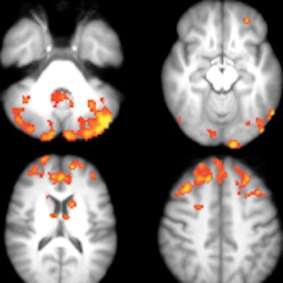
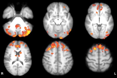
As one might expect, baseline resting-state fMRI showed that MS patients had significantly lower functional connectivity clusters in the cerebellum, frontal and occipital cortices, caudate nucleus, and thalamus than the healthy subjects.
 Axial statistical maps show areas of reduced thalamic functional connectivity in patients with MS compared with healthy subjects. Patients exhibited significantly lower functional connectivity in clusters located in the cerebellum, frontal and occipital cortices, caudate nucleus, and thalamus, bilaterally. Image courtesy of Radiology.
However, there was no significant difference in baseline functional connectivity maps of the thalamic resting-state network between the waitlist and intervention groups. The scans covered the cerebellum, thalamus, basal ganglia, and cingulate, frontal, temporal, occipital, and bilateral parietal cortices.
After the eight-week rehabilitation program, patients in the intervention group exhibited functional connectivity changes that correlated with improvements in sustained attention and executive function. By comparison, patients in the waitlist group, who did not participate in the video game training, showed no such changes.
| Cognitive test results from video game rehabilitation | |||
| Group | Baseline | After 8 weeks | p-value |
| Intervention | |||
| PASAT | 35.5 (± 10.1) | 46.4 (± 7.2) | 0.03 |
| SDMT | 37.5 (± 9.5) | 50.5 (± 17.9) | 0.013 |
| Stroop | 22.8 (± 4.9) | 28.8 (± 4.9) | 0.02 |
| Waitlist | |||
| PASAT | 32.2 (± 16.6) | 37.0 (± 10.9) | 0.127 |
| SDMT | 33.9 (± 8.6) | 39.0 (± 12.6) | 0.08 |
| Stroop | 24.2 (± 5.5) | 24.9 (± 8.1) | 0.65 |
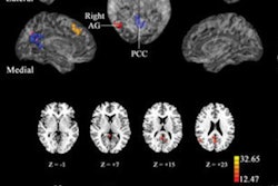
"The improvement in cognitive performance positively correlated with the functional connectivity increase in cortical areas belonging to the posterior components of the default mode network and negatively correlated with the functional connectivity decrease in cerebellar areas," the authors wrote.
The altered connectivity indicates that the gaming sessions changed the mode of operation in certain brain regions, De Giglio said in a statement released by RSNA.
"This means that even a widespread and common use tool like video games can promote brain plasticity and can aid in cognitive rehabilitation for people with neurological diseases, such as multiple sclerosis," she said.
The researchers cited several limitations of the study, including the focus on thalamic functional connectivity, which precluded the identification of changes in other resting-state networks.
"However, the relevance of thalamic regulation of the brain circuits involved in cognitive processes largely justifies our choice," they wrote.
The authors also cited the small patient sample and the exclusion of a healthy control group who underwent the same video game-based cognitive training.


