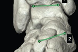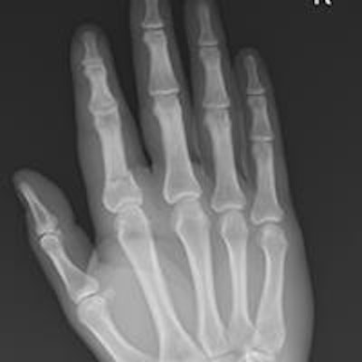
To help in daily practice, researchers from Portugal have conducted an in-depth assessment of the most important measurements used for upper and lower limb radiography examinations. They presented their findings at the 2015 congress of the European Society of Musculoskeletal Radiology (ESSR).
"Although sometimes overlooked, conventional radiography often provides important evidence of diagnostic value," noted the group from Centro Hospitalar Do Algarve in Portimao. "Using the available tools, namely simple and quick radiographic measurements, the radiologist should be able to provide straight answers in several clinical situations regarding the musculoskeletal system of the upper and lower limbs."
X-ray is usually the first imaging modality used to study the musculoskeletal system, because of its widespread availability, quick access, and low cost, but its interpretation isn't always straightforward and radiologists must use every tool available for the best possible examination, according to lead authors Dr. Carlos Bilreiro and José Saraiva. There are multiple measurements for upper and lower limb radiographic evaluations, which provide diagnostic criteria for numerous disorders.
In two poster presentations at the 2015 ESSR, they provided a clear depiction of the correct application of these measurements (see tables).
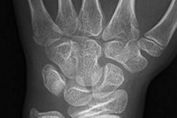
| Radial height | |
| Projection | AP view of the wrist |
| Purpose | Evaluate shortening of the radiue (e.g., after a fracture of the distal radius) |
| Technique | Distance between two lines perpendicular to the long axis of the radius: one line passing through the distal edge of the radial styloid and the other passing through the ulnar border of the radius |
| Criteria | Mean: 13.5 ± 3.8 mm |
| Comments | Despite the mean values reported, a comparison with the healthy side should always be done for a correct evaluation. A loss of radial height is associated with worsened outcomes. |
For lower limb radiographic evaluations, they assessed measurements for the hip, knee, ankle, and foot joints.
For instance, anteroposterior full-length standing radiographs often are used to evaluate the alignment of the lower limbs. The mechanical axis passes through the center of the femoral head and through the center of the ankle joint (midpoint of the tibial plafond), and the normal mechanical axis should pass just medial to the center point of the knee joint. The anatomic axis is defined by the axis of the femoral and tibial shafts.
The mechanical axis deviation (MAD) should be of 10 mm ± 7 mm. Genu valgum (knock-knee deformity) can be diagnosed when MAD is laterally deviated for > 3 mm (normal - standard deviation [SD]), and this condition should be considered when MAD is medially deviated for > 17 mm (normal + SD). The anatomical tibiofemoral angle is of 6.85° ± 1.4°. Genu valgum must be considered when the angle is higher than 8.3°, and genu varum should be considered when it is lower than 0°.
| Shoulder, acromiohumeral distance | |
| Projection | AP view of the shoulder (neutral) |
| Purpose | Diagnosing chronic massive rotator cuff tears |
| Technique | Distance between the inferior surface of the acromium and the humeral head |
| Criteria | > 7 mm - normal range < 7 mm - diminished |
| Comments | A shortening of the distance between the inferior surface of the acromium and the humeral head was found to correlate well with rotator cuff tears. |
"The most important image quality criterion for these measurements is to have the patellae centered between the femoral condyles," the authors wrote.
To evaluate lower limb length (relative and absolute), first draw a horizontal tangent line for the superior margin of the femoral head and then measure the length of the femur between that point and the horizontal tangent to the most distal point of the medial femoral condyle, they recommend. Tibial length is measured from the later tangent to the horizontal tangent through the center of the tibial plafond. Total limb length can be determined by measuring the total length from the superior border of the femoral head to the center of the tibial plafond.
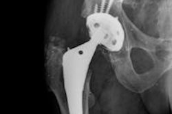
Anteroposterior pelvic or hip and femur radiographs are used to evaluate hip alignment and the important aspect is the angle formed by the longitudinal axes of the neck and shaft of the femur, according to Bilreiro and Saraiva.
| Elbow, cubital angle (humerus-elbow-wrist angle of Oppenheim) | |
| Projection | AP radiograph of the arm with the elbow in full extension and supination |
| Purpose | Diagnosing cubitus varus/valgus |
| Technique | Angle between the humerus and the forearm axes |
| Criteria | < 5° - cubitus varus > 15° - cubitus valgus |
| Comments | Also known as the elbow carrying angle, is normally slightly valgus, preventing the forearm from hitting the legs when walking. However, there is significant variation of the cubital angle between individuals, so a comparison with the healthy side should always be done to correctly assess the presence of a pathological angulation. |
At this year's ECR, the same authors presented new research about the range of imaging features of portal biliopathy, using a multimodality approach with pathologic correlation. They focused on the key imaging points necessary for a correct diagnosis.
"Portal biliopathy is a diagnosis that should be held in consideration in every case of biliary duct dilatation, most importantly when signs of portal vein cavernomatous transformation are present," they explained. "Although usually asymptomatic, associated cholestasis may lead to stone formation. A large range of conditions is included in the differential diagnosis, but even so, radiology is able to achieve the correct diagnosis."
Portal biliopathy represents the biliary repercussion of chronic portal hypertension, more frequently caused by thrombosis with cavernomatous transformation of the extra-hepatic portal vein, Bilreiro and Saraiva continued. It is usually asymptomatic and manifested by bile duct dilatation, localized or diffuse irregularities of the ducts contours, and focal narrowing or dilatation of the common and hepatic ducts. Additional manifestations may develop in rare cases of late diagnosis, such as associated choledocolithiasis and hepatolithiasis.
To view the full ESSR poster about the lower limb free of charge via the European Society of Radiology's (ESR) electronic poster system, EPOS, click here. For the e-poster about the upper limb, click here.


