A new sparse sampling MRI technique promises to speed up left ventricular (LV) volume and function assessments compared with the gold standard -- and to do so more accurately, according to a Swiss presentation at this month's European Society of Cardiology (ESC 2014) in Barcelona, Spain.
Results in 34 patients and volunteers showed that the compressed sensing (CS) technique was not only faster but also significantly more accurate and less variable than the conventional cine MRI approach for LV stroke volume assessment, which requires multiple breath-holds for the 10- to 15-minute acquisition.
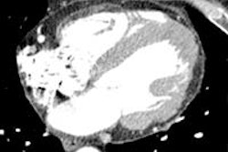
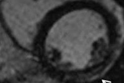
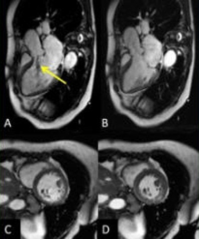 Above, compressed sensing acquisition in a 79-year-old with LV systolic dysfunction (LVEF 41%) and severe aortic regurgitation (regurgitation fraction 36%, arrow) performed before an aortic valve replacement. Images A and B represent a three-chamber view in diastole. C and D represent a mid short-axis view in diastole/systole. Neither flow-related artifacts nor fold-over artifacts can be seen. All images courtesy of Dr. Juerg Schwitter.
Meanwhile, high image quality was maintained in the sparse sampling technique, which will continue to evolve with further studies, according to Dr. Juerg Schwitter, director of the Cardiac MR (CMR) Center and associate professor at University Hospital Lausanne, Centre Hospitalier Univérsitaire Vaudois, and colleagues, who concluded that accurate and reproducible measurements of LV volumes and function can be obtained in a single breath-hold using the multislice CS cine MR sequences.
The technique provides for significant reductions in scan times as well, though the prototype system still needs refinement, the group reported.
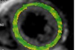
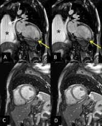 Compressed sensing acquisition in a 58-year-old patient with dilated cardiomyopathy, LVEF of 8%, an apical thrombus (yellow arrow), and pleural effusion (asterisk). A mild fold-over artifact can be seen in the two-chamber view (white arrow), but quality is sufficient to detect the apical thrombus. A/B represent a two-chamber view in diastole/systole. C/D represent a mid short-axis view in diastole/systole.
"We wanted first to test the whole concept in a study in volunteers and patients. Now, as we know the potential of the method, such technical improvements will certainly follow," Schwitter told AuntMinnieEurope.com in an email.
Three separate components are crucial for the concept of compressed sensing to work, he explained. "The acquired data must be sparse in order to guarantee compressibility of the input data (e.g., sparse in the time domain as applied in the presented cine heart imaging). The undersampling of data must be incoherent, so that artifacts (caused by undersampling) appear as noise. Finally, the reconstruction uses a nonlinear iterative optimization procedure to eliminate this noise from the images."
The study team implemented its single breath-hold multislice CS cine sequence on a 1.5-tesla MR system (Magnetom Aera, Siemens Healthcare), acquiring three long-axis and four short-axis slices over 14 heartbeats in a single breath-hold. Temporal and spatial resolution was 30 msec and 1.5 x 1.5 mm2, respectively. Data were analyzed using Argus 4DVF software (Siemens Healthcare).
To evaluate LV stroke volume, the authors measured aortic flow using a phase-contrast acquisition in 16 subjects who consisted of volunteers and patients without mitral insufficiency on echocardiography. Finally, they evaluated image quality of the compressed sensing sequences and the intra- and interobserver reproducibility.
The results showed that compressed sensing was more accurate than the conventional approach for quantifying LV stroke volumes.
| Results of single breath-hold compressed sensing cine images versus standard approach | |||
| Evaluation method | Stroke volume overestimation versus aortic flow | Variability of results | Quantitative accuracy in LV systolic function |
| Compressed sensing cine MRI | 6.4 ± 6.9 mL | r = 0.91 | CS-LVEF = 48.5 ± 15.9% |
| Conventional cine MRI approach | 14.1 ± 11.2 mL | r = 0.79 | standard LVEF = 49.8 ± 15.8%, (p = 0.11) |
The CS acquisitions maintained high image quality in 94% of subjects, maintaining qualitative accuracy in LV systolic function (above) and with excellent correlation (r = 0.96, slope = 0.97, p < 0.00001). Finally, intra-/interobserver agreement for all CS parameters was good (slopes: 0.93-1.06, r: 0.90-0.99), the group reported.
Accurate and reproducible measurements of LV volumes and function can be obtained in a single breath-hold using the new multislice compressed sensing cine sequence, with significant reductions in scan time and potential clinical application, the authors concluded.
"The beauty of the CMR techniques is that you can adjust several imaging parameters to best fit the patients' condition and the image quality you need," Schwitter told AuntMinnieEurope.com. "Thus, also in the case of compressed sensing you can adjust as needed, but compressed sensing allows to shorten drastically the breath-hold time (e.g., down to 1-2 heartbeats if you acquire a cine movie of one slice orientation only) or you take a breath-hold of 16 heartbeats to acquire as many slices as needed to reconstruct the entire left ventricle in 3D as we did in our study."
Reconstruction times are still substantial, but can be shortened considerably if technical improvements are made, such as several microprocessors used in parallel. Technical improvements will follow to fine-tune the acquisition process, he noted.
This project is really a proof of concept, he said, and its success means the technique can potentially be applied to many other CMR applications, such as tissue characterization to improve performance of coronary MR angiography, and perfusion acquisition to detect or exclude myocardial ischemia -- both of which are currently being tested at his facility, Jurg noted. Blood measurement and MR angiography acquisitions are also promising candidates for compressed sensing applications.
"Magnetic resonance of other nuclei such as C-13 carbon and F-19 fluorine allow us to assess metabolism in real-time and cell tracking in vivo, respectively -- we are working on these techniques with very encouraging results," Schwitter wrote. "As these novel MR techniques offer signal sparsity, they are ideal to benefit from this compressed sensing technique, as well. Indeed, MR imaging may go into a next phase of acceleration which opens new unexpected opportunities."

