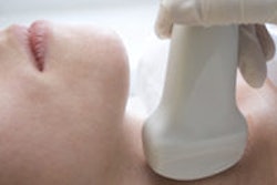
Ultrasound elastography has established a role in breast imaging, and the thyroid is one of the next clinical applications on the horizon. Polish researchers have unveiled a new data analysis method that may improve the technique's ability to characterize benign from malignant thyroid nodules.
Elastography is often based on the assumption that tissues deform in a linear fashion, and that changes in this deformation, or strain, can be signs of malignancy. But most soft tissues don't deform in a linear way, and this discrepancy calls for new ways to interrogate data.
In their presentation at the 2013 American Institute of Ultrasound in Medicine (AIUM) meeting, the researchers discussed how a nonlinear measure could significantly bolster elastography's performance in the thyroid. A team led by Dr. Rafal Slapa of the Medical University of Warsaw found that analysis of thyroid elastography time-strain curves led to 100% sensitivity and 85% specificity for characterizing thyroid nodules, a vast improvement over traditional elastographic analysis methods.
"The analysis of linear and nonlinear elastosonographic data may greatly improve differential diagnosis of thyroid nodules," Slapa said during his talk at a scientific session at the New York City meeting.
Although elastography can assist in the differential diagnosis of thyroid nodules, its diagnostic performance at present is not ideal, Slapa said.
"Further improvements in the technique and the diagnostic criteria are necessary for this exam to provide a useful contribution to diagnosis," he said.
Seeking to evaluate a new linear and nonlinear approach for interpreting thyroid strain elastography exams based on the evaluation of time-strain curves, the researchers studied 67 patients who were scheduled for thyroidectomy. All patients received B-mode and power Doppler ultrasound of the whole thyroid and neck lymph node.
During the ultrasound exam, 96 dominant thyroid nodules were examined using strain elastography on an Aplio XG ultrasound scanner (Toshiba Medical Systems) with a 5- to 17-MHz transducer. The nodules were evaluated using the classic method of qualitative analysis via elasticity scores and semiquantitative analysis with thyroid tissue strain/nodule ratios using the Elasto Q software (Toshiba). They also tested the new approach of evaluating time-strain curves on Elasto Q.
Of the 96 nodules in the study, seven were papillary carcinomas and 89 were benign. The classic elastography analysis methods of elasticity score and elasticity ratio did not show a significant difference between malignant and benign nodules (p = 0.431 and 0.156, respectively). However, a new parameter -- the relative length of nonlinear relaxation -- yielded excellent differentiation (p = 5.6 x 10⁻⁹) during the evaluation of time-strain curves, Slapa said.
A relative length threshold of 0.5 of nonlinear relaxation yielded 100% sensitivity and 85.4% specificity, with an area under the receiver operating characteristic (ROC) curve of 0.975.
The researchers acknowledged the limitations of their study, including the small number of malignant masses. In addition, the only malignant lesions found were papillary carcinomas.
Also, the strain/time ratio method is subject to the choice and number of images used for analysis, Slapa said.
Nonetheless, the findings show that the new approach has the potential to greatly improve differential diagnosis of thyroid nodules, he said.
"Further multicenter large-scale studies evaluating the usefulness of linear/nonlinear elastosonographic phenomena in differential diagnosis of thyroid cancer are warranted," he said. "Studies involving evaluation of viscoelasticity -- for example, shear-wave spectroscopy -- could be applied to differentiation of thyroid nodules."


