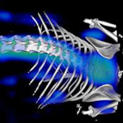
Datasets in optical and nuclear imaging are being fused with those from other modalities to provide better anatomical information. And in addition to merging PET or SPECT with MRI or CT, the combination of fluorescence molecular tomography (FMT) with CT or MRI is showing particular promise, a leading German expert said.
"Multimodal image datasets can be obtained from real hybrid imaging scanners or from fusion of separately acquired datasets," noted Dr. Fabian Kiessling, professor and chair of Experimental Molecular Imaging at Rheinisch-Westfälische Technische Hochschule (RWTH)-Aachen University.
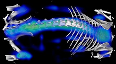 Merged fluorescence molecular tomography and µCT image of a mouse after injection of the targeted fluorescent imaging agent Osteosense. The agent targets hydroxyapatite to enable in vivo detection and monitoring of skeletal changes that occur during bone growth or resorption. Image courtesy of Dr. Fabian Kiessling.
Along with Dr. Bernd Pichler, a professor in the Laboratory for Preclinical Imaging and Imaging Technology of the Werner Siemens Foundation at Tübingen University Hospital, Germany, Kiessling is running a workshop about hybrid imaging at the European Molecular Imaging Meeting (EMIM 2013), which begins on 26 May in Turin, Italy.
"We will discuss how imaging data deriving from different modalities can be accurately merged," Kiessling explained. "A session will be dedicated to marker, surface, and intensity-based coregistration, including rigid and nonrigid surfaces. We will also address how image reconstruction and quantification can benefit from a hybrid imaging approach. This will include a discussion of how information from MRI and CT can be used to improve image reconstruction in FMT at the raw data level, and how CT and MRI can be used to perform attenuation correction in hybrid imaging devices."
There will be a critical discussion about the indications for hybrid image acquisition versus subsequent scanning approaches, and the workshop will end with a 30-minute video demonstration of a typical hybrid imaging experiment, he added.
 Information from MRI and CT can help to improve image reconstruction in fluorescence molecular tomography, according to Dr. Fabian Kiessling.
Over three days, EMIM 2013 will bring together scientists working in the diverse fields of molecular imaging and provide a platform for knowledge exchange between the disciplines, generations, and societies. It will help to strengthen the interaction between the various groups and everybody who is sharing a vision that interdisciplinary knowledge exchange is the basis for innovation, the organizers state on the EMIM 2013 website.
"The general aim has been to provide you with a concept which differs from the WMIC [World Medical Imaging Congress] to avoid repetition and to offer a workshop character," the organizers wrote. "We hope also to attract scientists from related fields, not only to the EMIM but also to the broad field of molecular imaging and to provide you with a broader view."
The plenary lectures will focus on functional and molecular imaging of the heart, imaging neural networks at work, imaging the tumor microenvironment, harnessing imaging to achieve controlled drug delivery to tumors, and intraoperative fluorescence imaging in cancer.
Overall, EMIM 2013 aims to provide a platform to further establish a sustainable and powerful European molecular imaging community.
"Close collaboration with the industry is most desirable to ensure the transfer of most recent knowledge. We need your support to build up a successful Europe-wide infrastructure in the field of molecular imaging and to organize a meeting where the surrounding conditions as well as the scientific level are excellent," stated the organizers.

