New iterative reconstruction algorithms are a considerable advance in musculoskeletal (MSK) CT, and allow a dose reduction of 50% and better imaging of metallic implants, according to award-winning research from France.
"Iterative reconstruction is very interesting for bone and soft-tissue analysis when metal hardware is present," noted Dr. Alban Gervaise, from the Service d'imagerie médicale, hôpital d'instruction des armées Legouest, Metz, and the Service d'imagerie Guilloz, hôpital central, CHU de Nancy, and France. "Traditionally, better visualization of metallic materials requires an increase of parameters such as the kVp and the mAs, as well as a low pitch and a thin collimation. All these parametric changes are a source of dose increase. Iterative reconstruction reduces the metal and streaks artifacts while avoiding a dose increase due to the optimization of the acquisition parameters."
Limiting the scan coverage to the zone of interest and narrowing the width of the scanned volume are simple ways to reduce the dose in MSK CT examinations, and iterative reconstruction must also be used whenever possible, he stated in an e-poster that was awarded a certificate of merit at RSNA 2012.
"The optimization of both tube current and tube potential remains a critical issue; these factors must be adapted to the type of exploration and the body habitus of the patient," Gervaise advocated.
Tube voltage must be adapted to the localization of the joint being scanned, the habitus of the patient, and the type of acquisition, with a reduction being made in the case of CT arthrography or vascular acquisition. In addition, tube current must be adapted to the indication (soft-tissue analysis needs higher tube current values than bone analysis), as well as localization of the joint being scanned and the habitus of the patient, he explained. During dynamic or perfusion CT, dose can be reduced by low kV acquisition and limitation of the scan coverage and the number of phases.
In clinical practice, justification and substitution also remain prime considerations. Behavioral aspects, such as respecting the indications, are essential, as is substitution, because ultrasound and/or MRI are often an option, particularly in MSK imaging, he added.
MSK CT scans are performed to evaluate different types of anatomical structures with different sizes (peripheral versus proximal joints) at locations with different radiosensitivity and tissue weight coefficient, and there are many different types of examinations, including standard CT, CT arthrography, vascular examination, and dynamic and perfusion CT.
The CT acquisition parameters are very different for each type of study; e.g., tube voltage setting ranges from 80 kV for a peripheral joint to 120 to 140 kV for the spine. Gervaise remarked that the doses are also very diverse and can vary by a factor of 1 to 1000; e.g., a low-dose wrist CT at an effective dose of 0.014 mSv compares with a standard dose lumbar spine CT at an effective dose of 14 mSv.
Weight tissue coefficients (k) are also very different for each anatomic region studied. The lumbar spine has a k of 11.2, which is quite similar to the abdomen, but the wrist has a k of only 0.22. Therefore, he thinks dose reduction is particularly important for spine and proximal joint CT examinations with high k factors.
Large dose variability can be explained by differences in z-axis coverage, which ranges from 5.8 to 25.5 cm. Another important factor is detector system design; multidetector CT doses are higher than single slice, but this is mainly due to an increase in z-axis scan coverage. Iterative or standard filtered back projection (FBP) reconstruction can also have an influence, as do the characteristics/body habitus of the patient population.
As a general rule on CT, the effective dose is lower the further you investigate from the body trunk, Gervaise noted. This is because peripheral joints are small in size, so tube output parameters and z-axis coverage can be shortened. Also, the tissue weight coefficient used to calculate the effective dose is very small.
"In MSK CT, most scans consist of a single-phase unenhanced acquisition. But, be careful with multiple phases performed during special conditions: interventional CT, perfusion CT, dynamic CT," he stated. "If it's not possible to reduce the number of phases, try to reduce the dose of these phases by limitation of the scan coverage and low dose acquisition."
A precise centering of the anatomical zone to be scanned at the isocenter of the CT gantry provides optimal image quality and delivered dose because more interpolations of the data are performed in the center of the gantry than in its periphery, he continued. The width of the scanned volume should be as narrow as possible to limit scattered radiation and beam-hardening artifacts.
During acquisitions of the lower limb, the contralateral limb should be flexed out of the scanning field when possible, while peripheral joints should be scanned as far as possible from the trunk of the patient in order to reduce the dose received in radiosensitive organs.
Reducing the tube potential (kV) can lower dose, but this also increases image noise. In practice, this increase in noise is not detrimental to analysis of bone structure due to its high natural contrast, according to Gervaise. Therefore, peripheral joints can be imaged at 80 kV. For larger proximal joints -- the shoulder, hip, sacroiliac, and spine -- the kV must be adapted to the body habitus of patients: 120 kV for a standard patient, 100 kV for thin patients, and 135-140 kV for larger patients.
Similarly, reducing the tube current (mA) causes an increase in image noise that can make it difficult to interpret examinations requiring a good contrast-to-noise ratio. The high natural contrast of bone structures allows low-dose acquisitions with considerable noise but with no effect on interpretation.
With some modern CT scanners, using the concept of effective mAs (mAs/pitch), pitch modification has no influence on dose because it is automatically adapted to the mA, he explained. To avoid motion artifacts and to follow the contrast bolus during vascular studies, the pitch factor should be adapted to the clinical conditions.
A high pitch of about 1.5 is preferred to reduce acquisition time and motion artifacts, e.g., for investigating a polytraumatized patient. The pitch should, however remain lower than 2 to keep an optimal quality of multiplanar reformations and to avoid helical artifacts. In contrast, a small pitch is preferred to reduce metal hardware-related artifacts.
In general, acquisitions are performed with thin slices (0.5-1 mm) to allow bone-structure analysis, and they are reconstructed in thicker slices (2-5 mm) for soft-tissue analysis. Submillimetric slices improve spatial resolution, reduce partial volume effects and allow the reconstruction in a quasi-isotropic volume. Thin slice acquisition can lead to an increase in image noise, so whereas acquisition is made in submillimetric slices, the soft-tissue analysis is performed on thick slices with a better signal-to-noise ratio.
Finally, to reduce the dose of dynamic and perfusion CT studies, he recommends reducing the mAs and the kVp respectively, as well as using iterative reconstruction, reducing the acquisition length, cutting the number of phases, and if possible, using volume mode instead of helical mode.
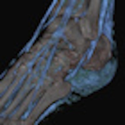
Editor's note: The CT image showing tendons in the foot that appears on our home page was provided by Dr. Thorsten R.C. Johnson, associate professor of radiology and head of CT, University of Munich, Grosshadern Campus.
