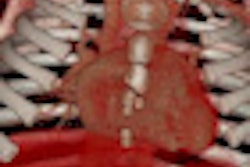
Getting images of sufficient quality for clinical evaluation in children as well as agitated or sedated patients represents a major challenge in abdominal MRI because the need for many sequences requires a breath-hold of about 15-20 seconds. Portuguese radiologists have outlined how to address this problem.
Respiratory triggering or navigation techniques are routinely incorporated in T2-weighted imaging in noncooperative patients, but the challenge is greater when it comes to avoiding breathing artifacts in T1-weighted (T1W) images. T1W images are of particular importance due to the acquisition of in-phase (IP) and out-of-phase (OP) images, and also their use in dynamic contrast studies, according to Dr. Filipe Veloso Gomes and colleagues from the radiology departments at Hospital Garcia de Orta and Hospital de Faro.
"MRI has become an essential technique in the evaluation of the abdomen in recent years. Although great technical improvements have been seen, motion remains, by far, the biggest difficulty in abdominal MRI," they noted in an e-poster at this month's European Society of MR in Medicine and Biology (ESMRMB) congress in Lisbon. "Recent advances in motion-resistant T1W abdominal imaging allows the evaluation of a full range of abdominal diseases in patients unable to cooperate with breath-hold."
Imaging noncooperative patients has been transformed by the use of a single-shot technique named magnetization-prepared gradient recalled echo (MP-GRE), as well as a radial acquisition technique called 3D gradient-recalled echo sequence with radial data sampling (radial 3D-GRE), the authors explained.
MP-GRE is an ultrafast GRE sequence using a small flip angle, very short repetition time (TR), and optimized K-space filling. In this setting, T1 contrast is preserved by preparing magnetization with a 180° inversion pulse, before the sequence repetitions ensue. K-space lines are acquired after a single inversion pulse (single-shot).
"The MP-GRE T1W, in-phase and out-of-phase, single-shot technique acquired in a free-breathing manner produces high-quality images in patients unable to cooperate," stated Veloso Gomes and colleagues. "T1W gradient-echo in-phase and out-of phase imaging is an essential component of comprehensive abdominal MR exams. It is useful for the study of fat-containing lesions and to identify various disease states related to the presence of fat in the liver."
Evaluation of this technique in the assessment of hepatic steatosis has been demonstrated, as well as the utility of this sequence for the characterization of adrenal lesions in noncooperative patients, they added.
3D-GRE with fat suppression is the method of choice for postcontrast image acquisition, but it is also vulnerable to breathing artifacts. Fat-suppression water excitation 2D-MPGRE is routinely used as a T1W postgadolinium fat-attenuated sequence in noncooperative patients.
Free-breathing 3D-GRE with radial data sampling has become established in thoracic and cardiac imaging, according to Veloso Gomes and colleagues. A preliminary evaluation of free-breathing 3D-GRE with radial data sampling acquisition in abdominal MRI has suggested that this technique is a feasible alternative to conventional 3D-GRE and with good image quality, particularly in noncooperative patients.
"Adequate image quality is usually achieved for T2-weighted imaging in noncooperative patients, either by the use of robust single-shot sequences or by the incorporation of respiratory triggering or navigation techniques," the authors explained. "T1W gradient-echo (GRE) IP/OP imaging is an essential component of comprehensive abdominal MR exams. Good image quality can be achieved for noncooperative patients by using a MP-GRE sequence technique."
Postcontrast studies in noncooperative patients can be performed effectively, with free-breathing 3D-GRE radial data sampling, they concluded.