MUNICH - Cardiovascular MR at 3 tesla is proving effective for detecting heart disease among cocaine addicts, Spanish researchers reported on Sunday at the European Society of Cardiology (ESC) annual meeting.
"Cocaine is a highly addictive drug with potentially lethal cardiovascular effects. The real prevalence and features of cocaine-related cardiovascular disease have been evaluated in selected groups only. We aimed to assess its prevalence and describe cardiovascular MR protocol at 3 tesla in consecutive cocaine addicts," noted lead author Dr. Alicia Maceira, from the cardiac imaging unit at the ERESA Clinic in Valencia.
A total of 94 patients who first attended a rehabilitation clinic were recruited for the study. One patient had a sudden cardiac death two days before undergoing MRI, and another had to be excluded when he was diagnosed with hypertrophic cardiomyopathy. Of the remaining 92 people, 13 were women and the average age was 37 (age range was 22-53 years).
 Between 14.3 million and 20.5 million people used cocaine in 2009, equating to between 0.3% and 0.5% of the global population ages 15-64, estimated the 2011 World Drug Report. Usage in Europe appears to have stabilized, however.
The mean duration of cocaine addiction was 7.8 years, and 78 patients were also tobacco smokers. Less than two months had passed between cocaine cessation and 3-tesla MRI. Only four patients were referred with mild cardiovascular symptoms, all of which involved palpitations, she stated.
End-diastolic volumes did not show differences with controls, according to Maceira, but end-systolic volumes were slightly enlarged (LV [left ventricle]: 31 ± 8 versus 24 ± 2 mL/m2, RV [right ventricle]: 38 ± 10 versus 28 ± 3 mL/m2, all p < 0.001) and so was the left ventricular mass index (80 ± 13 versus 69 ± 4 g/m2, p < 0.001). LV and RV ejection fraction (EF) were decreased compared with the controls (LV: 59 ± 6% versus 68 ± 4%, RV: 55 ± 5% versus 64 ± 6%, all p < 0.001).
About 35% of patients had decreased LVEF and 20% had decreased RVEF. About 29% of patients showed LV hypertrophy (14 concentric, 13 eccentric), and 12% presented concentric remodeling. No perfusion defects or necrosis patterns were seen, while 15 patients (26%) showed late gadolinium enhancement (one subendocardial, two subepicardial, 13 intramyocardial, and nine inferior ventricular junction).
True fast imaging with steady-state precession (true-FISP) cine sequences were used to measure left and right ventricular dimensions and EF, and short-tau inversion recovery (STIR) sequences were used in a short axis stack. A dipyridamole (0.84 mg/kg) myocardial perfusion study was performed with gadolinium-DTPA (0.1 mmol/kg). Inversion-recovery sequences were used for detection of late gadolinium enhancement and T2-weighted turbo spin-echo sequences were used for the thoracic aorta.
Images were analyzed by two independent observers, and the ventricular dimensions and function were compared with those of 60 gender- and age-matched healthy controls.
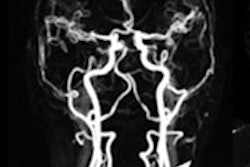
 Cardiac magnetic resonance end-diastolic images in the four- and two-chamber view of a subject with cocaine addiction. Concentric left ventricular hypertrophy can be seen, with a quantified left ventricular mass index of 103g/m². Images courtesy of Dr. Alicia Maceira.
"Cardiovascular MR at 3 tesla detected cardiovascular disease of variable degree in 69% of this cohort of consecutive cocaine abusers," Maceira concluded. "The main findings were a disease in systolic function of both left and right ventricles, an increase of left ventricular mass, and the presence of myocardial late gadolinium enhancement, suggestive of past myocarditis."
This is only a reasearch project and the authors are not suggesting 3-tesla MRI should be used routinely on addicts, she stressed.
In another study presented at the ESC congress on Monday, U.S. researchers found that in patients undergoing coronary CT angiography for acute chest pain in the emergency room (ER), those with cocaine abuse have a higher prevalence and volume of mixed coronary plaque than those without cocaine abuse.
Cocaine-associated chest pain accounts for a substantial proportion of ER visits in the U.S., according to Dr. Ullrich Ebersberger, from the Heart and Vascular Center at the Medical University of South Carolina. These patients are at increased risk of acute coronary syndrome and advanced atherosclerosis.
Ebersberger and his colleagues retrospectively studied 78 patients (52 men, mean age 44). The presence of stenosis was not significantly different between patients with and without cocaine use (12.8% versus 5.1% of patients, p > 0.05). Cocaine use was not associated with a higher prevalence of overall plaque (48.7% versus 35.9%, p > 0.05), calcified plaque (30.8% versus 21.8%, p > 0.05) or noncalcified plaque (16.7% versus 7.7%, p > 0.05).
In contrast, patients with a history of cocaine abuse had a higher prevalence of mixed plaque (34.6% versus 15.4%, p > 0.05) and mixed plaque volume (59.7 ± 33.3 mm3 versus 25.6 ± 12.6 mm3, p > 0.05).
"Although larger studies are needed, our results suggest that coronary plaque composition may play a role in the development of acute coronary syndrome in this patient population," Ebersberger stated.