An investigational raw-data-based iterative reconstruction scheme is getting high marks for image quality and dose reduction. Several research groups have compared the reconstruction technique with standard filtered back-projection (FBP) reconstructed images and uniformly found higher-quality images at around half the dose.
In a presentation at the 2012 European Congress of Radiology (ECR), French researchers stated clinical chest CT angiography images built with raw-data-based iterative reconstruction were better than traditional FBP images, even though they were acquired at half of the dose of FBP images. In another ECR 2012 lecture, a Dutch-led multinational effort showed reduced dose by 20 kV and still found less noise with iterative reconstruction. Finally, a study from Rotterdam compared FBP with raw-data-based iterative reconstruction in myocardial infarction patients and found better signal-to-noise ratio with iterative reconstruction and hence improved diagnostic confidence.
All three studies evaluated the Sinogram Affirmed Iterative Reconstruction (SAFIRE, Siemens Healthcare), an update on the firm's earlier-generation Iterative Reconstruction in Image Space (IRIS) technique. SAFIRE accesses raw data from the scanner rather than from image reconstructions as occurred in earlier iterative reconstruction schemes. Siemens is also offering new hardware that reduces reconstruction time. The SAFIRE technique is investigational in both the U.S. and Europe; 501(k) approval is pending in the U.S.
The group from Lille, France compared standard-dose filtered back projection (FBP) to low-Kv imaging with the SAFIRE algorithm in patients referred for follow-up chest CT; as a result all patients had a baseline CT scan at normal dose for comparison.
Iterative reconstruction offers a chance to provide high-quality images from low-dose datasets, said Dr. Julien Pagniez, from the department of thoracic imaging at Hôpital Calmette. "The purpose of our study was to compare the quality of CT angiography with 50% dose reduction using the raw-data-based iterative reconstruction algorithm SAFIRE by Siemens in comparison with standard filtered back-projection images," he noted.
"In daily practice, we set kilovoltage according to the body weight," said lead investigator Dr. Martine Rémy-Jardin, also from Lille. "Then for the follow-up there was a 20% kV reduction and we had to adapt the mAs because we reduced the kilovoltage, and it was done to obtain an overall 50% dose reduction ... What we wanted to test was the ability to restore image quality with iterative reconstruction."
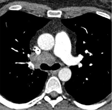 Chest CT exams obtained six months apart in a 40-year-old, obese woman (BMI: 36.5 kg/m2) referred for follow-up of thoracic malignancy. First scan performed with standard dose obtained at the level of the left pulmonary artery, and reconstructed using standard FBP (120 kVp DLP = 326 mGy.cm, Noise = 19.8 HU, and CNR = 8.1) delivers higher noise and radiation dose than low-dose SAFIRE reconstructed image from the same patient six months later, below (100 kVp DLP = 143 mGy.cm, Noise = 17.1 HU, and CNR = 9.2). Despite the dose reduction in the follow-up image, objective image noise measured at the level of the trachea was slightly reduced, and the contrast-to-noise ratio was slightly improved. Images courtesy of Dr. Julien Pagniez and Dr. Martine Rémy-Jardin.
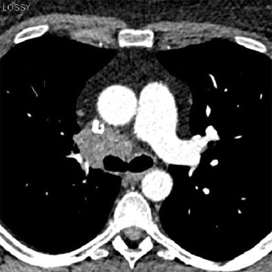
The 80 consecutive patients undergoing follow-up chest CT scans all had their second examinations acquired in a manner similar to the first scan, except that kV was reduced by 20% in the second scan to produce a 50% reduction in CT Dose Index Volume (CTDIvol). SAFIRE was used to reconstruct the images in the low-dose scan, Pagniez said.
In both sets of images, the authors obtained objective noise measurements by measuring the standard deviation of XL values in regions of interest in mediastinal images at two anatomic levels -- the tracheal lumen and the descending aorta -- and signal-to-noise ratios were calculated from these levels. In addition, the visual appearance of noise was rated as minimal, moderate, or severe, and the overall image quality was evaluated subjectively.
"There was an immediate reduction of 52.8% in the dose-length product," between the baseline T1 exam (163.6 mGy cm) and the follow-up T2 scan (77.3 mGy cm), Pagniez said. The results also showed significantly less objective noise in the trachea in mediastinal (T2: 18.33 ±5.89; T1: 21.48 ±9.41) (p < 0.0001) and the lung images (T2: 41.28±10.39; T1: 49.77±11.73) (p < 0.0001).
The low-dose protocol also demonstrated significantly higher signal-to-noise (T2: 12.92±3.80; T1: 10.86±3.15) and contrast-to-noise (T1: 11.68±3.64; T1: 9.42±3.13) ratios (p < 0.0001).
Finally, in subjective evaluations of image quality, the two thoracic radiologists evaluating the images found similar visual perception of noise on mediastinal (p = 0.1317) and lung images (p = 0.3657), mainly rated as moderate; and (d) similar overall image quality, rated as excellent in 23 % (18/80) (versus T1: 21/80; 26%) and good in 77 % (62/80) (T1: 59/80; 74%) of CT exams (p = 0.4054).
Overall image quality between the normal-dose and low-dose SAFIRE images was rated excellent in 23% of the cases, (18/80) (versus T1: 21/80; 26%), and good in 77% of cases (T1: 59/80; 74%) of CT examinations (p=0.4054), the group reported.
"Despite the dose reduction, there was a significant reduction in objective noise at the level of the trachea on both mediastinal and lung images on the follow-up lung images with low kV and iterative reconstruction," Pagniez said. "There was no significant difference in objective noise between the two groups at the level of the aorta on mediastinal images. The signal-to-noise and contrast-to-noise ratios were significantly improved at T2."
The technique provided equivalent image quality compared to low kV chest CT angiograms at a 50% dose reduction, according to Pagniez. "This suggests the possibility for reduced dose while improving vascular enhancement on low-dose chest CT exams in routine clinical practice."
In their study, Dr. Richard A.P. Takx, and colleagues from Maastricht University Medical Center in the Netherlands, along with investigators from the Medical University of South Carolina and two other centers, compared image noise between the raw-data-based reconstruction algorithm and traditional FBP. The study examined 20 consecutive patients who underwent coronary CT angiography using the pECGdual_step scan protocol.
"Second-generation DSCT hardware provides a prospective dual-step ECG pulsing (pECGdual_step) protocol in which adaptive prospective ECG-triggered cCTA acquisitions are combined with ECG-gated tube current modulation," Takx wrote in an email to AuntMinnieEurope.com. "Specifically, full tube current is triggered to a limited portion of the cardiac cycle (e.g., at 30% or 70% of R-R) for coronary artery analysis while low-radiation tube current (20% mAs) is applied during the majority of the cardiac cycle (e.g., 10-90% R-R), thus providing the reduced dose features of prospective triggering while obtaining a functional image series."
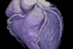
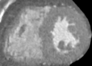 Short axis reconstruction of low dose (at 20% mAs) coronary CT angiography image reconstructed with standard FBP. In bottom image, low-dose CCTA image reconstructed with SAFIRE, demonstrates greatly improved image quality. Images courtesy of Dr. Richard Takx.
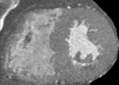
Takx and colleagues reconstructed the images using regular FBP at full-dose (Group 1a) and low-dose (Group 1b) and with SAFIRE with five iterations at low-dose (Group 2). They evaluated three regions of interest (ROI) for signal intensity, image noise, signal-to-noise-ratio (SNR) and contrast-to-noise-ratio (CNR). Finally, they evaluated subjective image quality on a four-point scale based on image sharpness and noise.
"Our objective was to compare objective and subjective image quality parameters using SAFIRE versus conventional FBP reconstructions in patients who underwent low-dose CCTA (coronary CT angiography)," Takx noted.
Objective image noise was reduced by an average of 22 % (P < 0.0001) in Group 2 compared to Group 1b. There was also a significant (P = 0.0156) improvement in overall subjective image quality in Group 2 compared to Group 1a.
Iterative reconstruction enabled significant reduction in image noise without loss of diagnostic information, and has the potential to improve image quality in low-dose functional CCTA and possibly reduce radiation dose even further, the group concluded.
"DSCT (dual-source CT) still remains sensitive to motion artifacts, especially in patients with arrhythmia," Takx stated. "In case of a motion artifact the images acquired in the low dose ECG-pulsing window can potentially be used for evaluation of coronary patency. However, these low dose FBP reconstructions contain a high level of image noise. In our study we demonstrate that low-dose CCTA reconstructed with the SAFIRE algorithm resulted in significant reductions in image noise and significant improvements in objective (SNR and CNR) and subjective image quality compared to low-dose FBP reconstructions."
Iterative reconstruction in myocardial infarction
Finally, a study from Erasmus University Medical Center used low-dose CCTA with SAFIRE iterative reconstruction in patients with a history of myocardial infarction. Dr. Alexia Rossi and colleagues scanned 19 patients with CCTA using a DSCT scanner (Siemens Healthcare), using prospectively triggered delayed-enhancement imaging performed eight minutes after CCTA. Images for each patient were reconstructed twice: first with FBP, and then using the SAFIRE algorithm at both 60% and 100% strength.
Signal and noise were measured in the descending aorta, and attenuation within the infracted myocardium was measured to calculate the contrast-to-noise (CNR) ratio, the authors wrote in an accompanying abstract. Two readers blinded to the results graded the images subjectively for image quality and diagnostic confidence in the detection infracted regions using a four-point scale ranging from 1 (poor) to 4 (excellent). MR delayed enhancement was used for comparison in all cases.
Compared to FBP, the use of SAFIRE at 60% strength reduced image noise by 26 ± 4% for the I26 kernel and 20±7% for the I36 kernel (p<0.001). Increasing the IR algorithm strength to 100% further reduced the noise to 39±9% for the I26 kernel, and 45±14% for the I36 kernel (p < 0.001). CNR also increased from 1.9±0.7 with the B26 kernel to 2.6±0.7 with the I26 kernel at 100% strength (p<0.05). "The overall image quality and the diagnostic confidence significantly improved using iterative reconstruction," the authors wrote.


