Functional imaging modalities such as fMRI identify regions of brain activity by measuring changes in blood flow. But while fMRI offers excellent depth penetration, its sensitivity and temporal resolution are limited.
Now, researchers at the Institut National de la Santé et de la Rechereche Médicale (INSERM) in Paris have come up with an alternative: functional ultrasound, an ultrasensitive imaging technique that can visualize whole-brain microvasculature dynamics with high spatiotemporal resolution (Nature Methods, August 2011, Vol. 8:8, pp. 662-664).
Previously, ultrasound's low sensitivity meant that it could only be used to image blood flow within major vessels. To address this limitation, the INSERM researchers developed a high-speed imaging technique that can visualize flow in tiny brain vessels, where subtle variations are linked to cerebral activity. They claim that this functional ultrasound (fUS) method can image transient changes in blood volume with better resolution and sensitivity than previous functional brain imaging modalities.
With conventional ultrasound, tissue is scanned line by line with a focused beam. With a typical frame rate of about 50 frames per second, this process is too slow to acquire large-field images at the kilohertz rate required to image blood dynamics. To speed things up, fUS employs plane-wave illumination, in which a plane wave scans the whole image in a single shot. This boosts the frame rate up to about 10,000 frames per second.
"This means that, compared to conventional ultrasound, the number of time samples available for each pixel of the image is typically 100 times greater," explained Mickaël Tanter, PhD, the research director at INSERM. "As we have many more time samples per pixel, we are able to detect much more subtle motion than in conventional ultrasound."
In vivo activity
Tanter and colleagues tested the effectiveness of this method on rats, by imaging the areas of the brain that activate in response to a stimulus. To perform fUS, the researchers acquired 200 compound images at 1 kHz. Each 2 x 2-cm compound image was summed from 17 planar illuminations tilted with different angles into the brain, a process that increases image resolution and reduces noise.
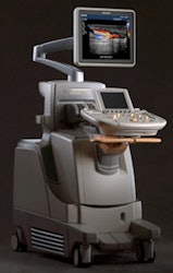
 Ultrasound has made rapid advances in recent years, and now the modality may be of increasing use in the brain. Image courtesy of Philips Healthcare.
Using a 15-MHz ultrasound probe, this method achieved a spatial resolution of 100 x 100 µm, a slice thickness of 200 µm, and a penetration depth of more than 2 cm -- sufficient to image an entire rat brain in just 200 msec. Tanter notes that this depth could be increased for human applications. "On adults during surgery, we could image the whole brain, typically up to 8 cm in depth," he said. "On newborns, we could image the whole brain noninvasively through the fontanel."
Tanter's co-authors Gabriel Montaldo, PhD, and Emilie Macé, PhD, measured power Doppler images (the mean intensity of the blood signal, which is proportional to the blood volume) every three seconds on rats during whisker stimulation. Whisker motion caused an increase in blood volume in the rat's primary somatosensory barrel cortex, indicating activity in this area. During stimulation, the power Doppler signal increased by between 10% and 20%.
Activation maps showing the correlation between signal and the temporal pattern of stimulus revealed significant activation of the somatosensory cortex during whisker motion, as well as significant activation in the ventral posterior medial nucleus. To further test fUS sensitivity, the team repeated the experiments while deflecting a single whisker to activate an individual cortical column. Maps from 10 trials showed significant activation of a small region in the somatosensory cortex.
The team also evaluated fUS for imaging transient brain activity, by examining four rats with induced epileptic seizures. They performed fUS acquisitions every 3 sec following focal injection of a potassium channel blocker. Videos of blood volume dynamics during the seizure revealed increasing blood volume at the injection site, followed by diffuse and bilateral spreading.
Different spreading patterns of blood volume changes were observed, including cortical waves crossing in both hemispheres, localized ipsilateral corticothalamic activations, or a wave traveling through the whole thalamus. The authors claim this is the first time that an epileptic seizure has been imaged in the whole brain at such spatiotemporal resolution.
fUS technology could open new avenues in neuroscience, according to Tanter. "For therapy, localizing the epileptic focus using fUS during surgery will be a major application," he told medicalphysicsweb. "Beyond clinical application, it will be a fantastic tool for people in neuroscience working on small animals. This technology should help them answer unsolved questions. We plan to miniaturize the probe for chronic awake animal studies and so provide a portable functional imaging system for neuroscientists."
"Functional imaging on newborns will also be of major interest in order to increase our knowledge in cognitive science," he continued. "We are implementing this mode to be compatible with pediatric ultrasonic probes for first human trials on newborns in 2012."
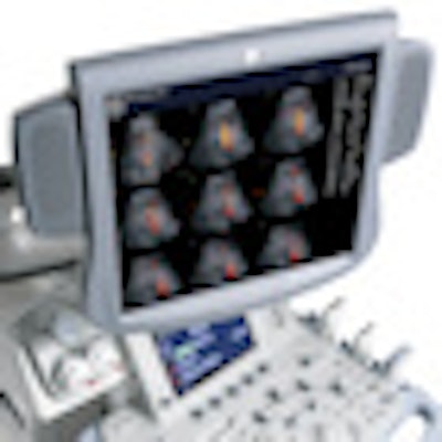
The photo on the home page is of the Logiq9 system. Image courtesy of GE Healthcare.
© IOP Publishing Limited. Republished with permission from medicalphysicsweb, a community website covering fundamental research and emerging technologies in medical imaging and radiation therapy.


