New technologies for mammography such as breast SPECT, positron emission mammography, and MR imaging show great promise for detecting and localizing breast cancer. In the past few years, traditional film-screen and digital mammography has benefited by the application of computer-aided detection (CAD) software.
Although studies have shown that the use of CAD can increase the detection of early-stage malignancies, radiologist experience is needed to reduce the number of false negatives to a minimum, according to an electronic poster presentation at this year's European Congress of Radiology (ECR) in Vienna, Austria.
A team from the Diagnostic and Interventional Radiology Institute at Maggiore della Carita Eastern Piedmont University in Novara, Italy, conducted research related to false-negative diagnosis with CAD. The group performed a retrospective analysis of 83 consecutive patients imaged at their facility from January 2003 to August 2004.
The patients ranged in age from 30 to 87 years, with a mean of 61.2 years. All patients had proven breast cancer that was assessed by ultrasound and mammographic imaging, and confirmed via histopathology.
The mammographic exams were conducted on a Senographe 2000D full-field digital mammography (GE Healthcare, Chalfont St. Giles, U.K.) unit that was integrated with a CAD system developed by R2 Technology (Sunnyvale, CA), stated Dr. Valentina Annibale, one of the study authors, in an e-mail to AuntMinnie.com.
All mammographic images were examined by the CAD application and by two radiologists experienced in mammography. The radiologists examined the images with consideration for presence of nodules, microcalcifications, and architectural distortion and reported according to the American College of Radiology's BI-RADS lexicon, the study authors wrote.
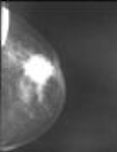
The researchers found that three of the 83 (4%) histologically proven breast cancers did not return a positive result in their CAD analysis in either the craniocaudal or mediolateral oblique views. In addition, they reported that in nine of the 83 patients, the CAD analysis showed the lesion in only one projection.
| Histologically proven breast cancer did not return a positive CAD result in either the craniocaudal or mediolateral oblique views. Image courtesy of Dr. Valentina Annibale. |
In the CAD false-negative group for both craniocaudal and mediolateral oblique projections, two of the three patients had a nodular lesion while the remaining patient presented with a parenchymal distortion. In the group with false-negative CAD in one of the two projections, four patients had a nodular lesion, four had nodular lesions with microcalcification, and one patient had both parenchymal distortion and microcalcification, according to the researchers.
The study team noted that the CAD system correctly returned positive results for both projections in 71 of the patients, and that they view the application as an important aid for mammography imaging. But because the false-negative examination rate for the application was 4% in their study, the researchers expressed concern with implicitly relying on CAD results.
"False-negative examinations represent a serious limit for its diagnostic accuracy," they wrote.
By Jonathan S. Batchelor
AuntMinnie.com staff writer
June 30, 2005
Related Reading
CARS panel discusses the future of CAD, June 28, 2005
CAD segmentation for mammo acceptable -- except in heterogeneously dense breasts, April 26, 2005
CAD offers promising second opinion, even in dense breasts, March 15, 2005
CAD systems show double-digit growth in Europe, November 23, 2004
CAD may unmask unsuspected cancers in diagnostic mammo, October 27, 2004
Copyright © 2005 AuntMinnie.com


