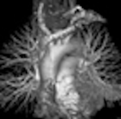
A new way to use CT angiography (CTA) to scan fast-moving targets -- such as fidgety children and grown-ups with rapid heartbeats -- is becoming routine at a hospital in France.
Patient management at the center has improved with the introduction of ultrahigh-resolution scans of the thorax, which are being used to find lung abnormalities as well as coronary artery disease without the need for breath-holding or electrocardiogram gating.
"What do we need for an optimal chest exam? The first thing is high spatial resolution because it's necessary to find even subtle abnormalities in the lung parenchyma," said Dr. Martine Rémy-Jardin, Ph.D. "We need high spatial resolution for evaluation of thoracic vessels. And we need high temporal resolution because ... the heart is beating and this is responsible for motion artifacts which can mimic abnormalities."
Rémy-Jardin, who heads the radiology department at University Center of Lille and thoracic imaging at Hôpital Calmette in Lille, France, discussed the high-pitch chest scans at Stanford University's International Symposium on Multidetector-Row CT in May.
Using a dual-source MDCT scanner, the thorax can be scanned at a temporal resolution of 83 msec, she said. The new-generation model (Somatom Definition Flash, Siemens Healthcare, Erlangen, Germany) that became available in 2009 further bumped up temporal resolution to 75 msec. The high temporal resolution stops motion artifacts even in fast hearts, while pitch values as high as 3.2 shorten the scan and reduce the dose.
"I'm sure this is going to be very interesting in clinical practice," Rémy-Jardin said.
To date, her group has performed a couple of studies using a prototype version of the Flash scanner (maximum temporal resolution, 83 msec; maximum pitch, 3).
Pitch 2 versus pitch 3
The first study, performed on the prototype Flash scanner with standard CTA protocols (80-140 kV, 90-120 mAs, 32 x 2 x 0.6 collimation) and tube-current modulation with Siemens CareDose 4, aimed to compare image quality at pitch 2 versus pitch 3. A total of 120 mL iodinated contrast at 35% was injected at 4 mL/sec prior to scanning, and 1-mm thick reconstructions were examined on a workstation.
Image quality was excellent for both pitch settings, with no statistically significant differences in image quality (p = 0.46). Vascular enhancement at the ascending aorta, descending aorta, and pulmonary trunk was 302.23, 289.47, and 306.06 for pitch 2, versus 313.09, 300.04, and 333.8, respectively, for pitch 3 scans.
The pitch 3 group had slightly higher noise, less motion artifact, and a lower radiation dose, Rémy-Jardin said.
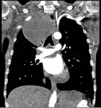 |
| A 47-year-old patient presented with shortness of breath. Ultrafast pitch 3 coronary CTA revealed a large mass in the trachea. The quality of the image acquired in 2.71 seconds was rated as excellent. All images courtesy of Dr. Martine Rémy-Jardin, Ph.D. |
"We found very good enhancement, despite the unsophisticated injection protocol," she said. "With pitch 3 we had an increase in objective noise, but this was not responsible for any degradation in the overall image quality or the diagnostic quality of the scans. And, of course, the higher the pitch, the shorter the acquisition time [mean 3.53 sec versus 2.54 for pitch 3] and the lower the dose-length product [DLP] value [mean DLP 228.1 mGy/cm versus DLP 176.9 mGy/cm for pitch 3, p < 0.0001], which means a lower dose."
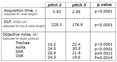 |
| In a comparison of 70 patients scanned at pitch 2 versus 70 at pitch 3, acquisition times and doses were lower at pitch 3. Image quality did not differ significantly between the two techniques, but objective noise was slightly higher using the pitch 3 protocol. Data from Tacelli, Rémy-Jardin et al, RNSA 2008. |
Pitch 3 only
A second study, completed in May by Demalherbe, Rémy-Jardin, and colleagues, scanned 93 patients without beta-blockers, electrocardiogram gating, or breath-holds. The scans covered the entire thorax in a mean of 1.75 seconds.
"For the pitch 3 protocol, we sought again to look at the coronary arteries because segments of the coronary arteries are of interest to radiologists for screening purposes," she said.
Considering the coronary arteries vessel-by-vessel based on four proximal segments per patient, on average, 3.5 (range, 1-4) were analyzable, Rémy-Jardin reported. Among the proximal and middle coronary artery segments, on average, 5.2 (range, 1-7) were analyzable. In all, 63.4% of the patients had four analyzable segments (59/93), while only 20.4% (19/93) of patients had seven segments analyzable due to the lack of electrocardiogram gating.
The mean dose-length product was 152.1 mGy/cm.
The overall image quality was excellent. Even in a patient with a heart rate of 82 beats per minute, "it is clearly possible to think of the coronary arteries at the screening level," even though it wasn't a dedicated cardiac exam, Rémy-Jardin said.
The introduction of the commercial model of the scanner this year has made it possible to acquire ultrafast high-pitch scans of the heart during the systolic phase, Rémy-Jardin said.
"This is going to be synonymous with improvement in the quality of diagnostic scans, and the diastolic phase will probably be the next area of analysis for the coronary arteries," she said.
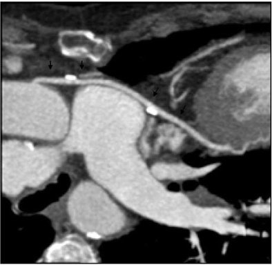 |
| Nongated ultrafast dual-source CT angiography (pitch 3) revealed sharp delineation of coronary artery bypass graft (CABG) at DLP of 134 mGy/cm, despite a high heart rate of 96 beats per minute. |
Moreover, by performing a diastolic acquisition immediately followed by a systolic acquisition of the coronary arteries, it has once again become possible to include a cardiac functional analysis in a thoracic scan -- a new way to think about cardiothoracic imaging, she said.
"High-pitch exams are interesting because we can get exams of excellent quality in clinical practice," Rémy-Jardin said. The technique is robust enough to have become standard protocol in the group's clinical practice, with experience in more than 500 cases.
The protocol is suitable for all patients, "but in particular those who cannot cooperate because they are too ill or too young," she said. "We can start examining patients who are breathing, and we can start obtaining examinations of children ... and patients under general anesthesia."
Additional benefits include very low doses and a low volume of contrast material.
By Eric Barnes
AuntMinnie.com staff writer
July 7, 2009
Related Reading
New lung cancer staging guidelines reflect improved CT, survival data, June 1, 2009
Triple-rule-out CT scans benefit from prospective gating, May 28, 2009
Tube current modulation cuts triple rule-out CTA dose, April 15, 2009
CT protocols reduce risk for pulmonary embolism, March 9, 2009
320-detector-row CT cuts dose in triple rule-out exams, February 12, 2009
Copyright © 2009 AuntMinnie.com


